Download
1 / 34

E N D
ANAEMIAS assoc.prof. T.Datsko
Anaemiais a blood disease of erythrocytes quantity or their hemoglobin saturation per unit blood volume. At the same time in the circulating blood there can appear erythrocytes of different sizes (poikilocytosias, poikilocythemia), different shapes (anisocytosis), different levels of colouring (hyperchromatism and hypochromatism), erythrocytes with inclusions (Jolly’s corpuscles, Kabo’s rings), nuclear erythrocytes (erythroblasts, normoblasts, megaloblasts).
To define the peculiarities of anaemia, morphogenesis and other blood diseases, biopsy of the sternal bone marrow puncture is widely used. In breast bone (sternum) punctate it is possible to diagnose the bone marrow regeneration level in anaemia as well as the type of erythropoiesis (erythroblastic, normoblastic, megaloblastic).
According to the etiology and pathogenesis, there are three groups of anaemias: posthemorrhagicanaemia (as a result of blood loss), anaemia as a result of erythropoiesis disturbance, and hemolytic anaemia (as a result of increased haemolysis). According to the clinical course anaemia can be acute and chronic. Classification of anaemias
Posthemorrhagicanaemia develops as a result of massive hemorrhage of the stomach or intestinal vessels due to ulcer or tumor effects, uterine tube rupture in extrauterine pregnancy, rupture of the aorta, pulmonary vessels disturbance in tuberculosis, etc. Because of the bleeding of large vessels the acute posthemorrhagicanaemia occurs and death occurs faster than morphologic manifestations of anaemia.
Because of the prolonged bleeding of small vessels the chronicposthemorrhagicanaemia develops and its manifestation can be pallor of the skin, mucous tunics, and viscera. Hyperplasia of the red bone marrow of flat bones and epiphysial plates turning intense and succulent. Metaplasia of yellow bone marrow occurs, turning red, the centres of extramedullary erythropoiesis in the spleen, thymus, lymph nodes and other tissues occurs. As a result of hypoxia (oxygen starvation) dystrophic changes occurs in the viscera, small hemorrhages in mucous and serous tunics may develop.
Sinusoidal megakaryocytes, lymph node Sinusoidal hematopoiesis, spleen
Anaemia as a result of erythropoiesis disturbance develops due to the deficiency of iron, vitamin B-12 and folic acid. Examples of this are hypoplastic and aplastic anaemiae. Asiderotic (iron-deficiency) anaemia is always hypochromic and develops as a result of low intake of iron into the organism with food. Such anaemiae are common among children, and also under intense need of iron during pregnancy, female maturation(from puberty to about 30 years) or chlorosis. This anaemia can appear in stomach and intestinal diseases, especially after their resection. Anaemia as a result of erythropoiesis disturbance
Vitamin B12 and folic acid deficiency anaemias (megaloblastichyperchromatism, pernicious (Biermer's, Biermer-Ehrlich) anaemia) are characterized by erythropoiesis disturbance and appear in disturbance of thaabsorbtion of exogenous vitamin B-12 in the stomach, in diseases of the stomach, with decreased secretion of gastromucoprotein. Such changes can be of hereditary origin or autoimmune genesis.Atlymphogranulomatosis, polyposis, syphilis, corrosive (necrotic, (toxico)chemical) gastritis, malignant growths of stomach, after the ulcer of the stomach, intestinal resections pernicious anaemia can appear. The cause of such anaemia can be deficiency of exogenous vitamin B-12 or folic acid of children fed with goat's milk. As a result of this the erythropoiesis is realized by the megaloblastic type and the hemolysis exceeds the erythropoiesis. Vitamin B12 and folic acid deficiency anaemias
The pathomorphologic manifestations of this anaemia are as follows: liver, spleen, kidney hemosiderosis, fatty degeneration of parenchymatous organs, general obesity, bleach lemon-tinged skin, small hemorrhages in mucous and serous tunics and in the skin. In the gastrointestinal tract, there are atrophic and sclerotic changes, the bone marrow turns raspberry-red with the predominance of erythroblasts, normoblasts, and megakaryoblasts. In lateral and posterior (dorsal) columns of spinal cord there is funicular myelosis and in the brain there are the centres of encephalomalacia and ischemia.
Hypoplastic and aplastic anaemias can be endogenous or inherited (familial aplastic anaemia of Fanconi and Ehrlich’s hypoplasticanaemia), and exogenous or acquired (radiation, toxic, medicamentosisanaemias). Hypoplastic and aplastic anaemias
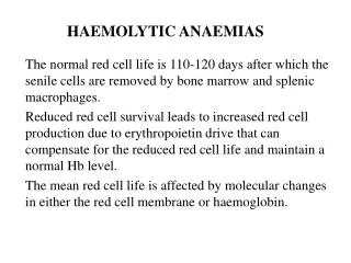
Hemolytic anaemia is characterized by the increased haemolysis which can be intravascular and extravascular. Intravascular anaemia appears when hemolytic poisons get into the organism, in bad burns (toxic anaemia), in malaria, sepsis and other infections (infectious anaemia), blood transfusion of incompatible blood group or Rhesus factor (posttransfusionanaemia), at immune pathologic processes (immune, isoimmune and autoimmune anaemias (hemolytic disease of newborns, chronic lympholeukemia, bone marrow carcinomatosis, systemic lupus erythematosus, medicamentosis immune hemolysis, thermal hemoglobinuria and other). Extravascular (intracellular) anaemia is mostly of inherited origin and is divided into erythrocytopathy, erythrocyte-enzymopathy and hemoglobinopathy. Hemolytic anaemia
Diseases such as microspherocytosis, inherited ovalocytosis, etc result in hemolytic anaemia due to their deviation from normal structures of the erythrocytes’ membrane. Erythrocyte-enzymopathic hemolytic anaemia appears due to deficiency of enzymes of pentose-phosphate cycle - glucose 6-phosphate dehydrogenase and pyruvate kinase. This anaemia grows progressively worse in viral infections, usage of some medicaments. Hemoglobinopathic hemolytic anaemia develops in disturbance of haemoglobin synthesis – a and b-thalassemia or in appearance of anomalous haemoglobin – S, C, D, E. Falciform cellular anaemia can include hemoglobinopathies.
Morphologic manifestations of hemolytic anaemias are very specific: general hemosiderosis, hemolytic jaundice in serious cases with hemoglobinuricnephrosis, splenomegaly in inherited hemolytic anaemias, the presence of centres of extramedullar erythropoiesis.
In contrast to aplastic anemia, leukemia results in a highly cellular marrow. The marrow between the pink bone trabeculae seen here is nearly 100% cellular, and it consists of leukemic cells of acute lymphocytic leukemia (ALL) that have virtually replaced or suppressed normal hematopoiesis. Thus, though the marrow is quite cellular, there can be peripheral cytopenias. This explains the complications of infection (lack of normal leukocytes), hemorrhage (lack of platelets), and anemia (lack of red blood cells) that often appear with leukemia.
Thrombocyte diseases. Diseases which manifest themselves in reduced quantity of platelets in circulating blood as a result of their increased destruction or decreased production are called thrombocytopenias. They can be inherited or acquired. Inherited thrombocytopenias are divided into immune and non-immune. Immune thrombocytopenia appears in incompatibility of blood in any system, in the disturbance of antigenic thrombocytes structure (heteroimmune), in production of antybodies against their own thrombocytes (autoimmune). Non-immune thrombocytopenia appears in case of mechanic injuries of thrombocytes, impaired proliferation of bone marrow cells because of toxic agents, radiation, metastases of malignant growths, hemoblastosis, vitamin B-12 or folic acid deficiency, disseminated intravascular coagulation (DIC), etc. Morphologic manifestation of thrombocytopenia is the presence of hemorrhagic syndrome on the skin, mucous tunics, and parenchyma of internal organs. Thrombocyte diseases
Thrombocytopathies are diseases in which morphologic, functional, biochemical thrombocytes impairments is observed, which causes the hemorrhagic syndrome development in the vessels of microcirculatory channels. Thrombocytopathies can be congenital or acquired. They are characterized by the disturbance of the formation of hemostatic thrombocyte plug including adhesion, secretion, and aggregation. Inherited variants of pathology mostly accompany other inherited defects. In their essence there is autosomal recessive disturbance of membrane glycoprotein synthesis and thrombocytes secretion. As an example we can observe Glanzmann -Negeli(Glanzmann’sthrombasthenia) disease with lack of thrombocytes aggregation, the disturbance of binding with fibrinogen and prolonged bleedings. The other example is Bernard-Soulier syndrome with large thrombocytes and reduction of their adhesion. Acquiredthrombocytopathies appear in many diseases: hemoblastosis, vitamin B-12 deficiency anaemia, cirrhosis, tumour diseases of the liver, uraemia, radiation sickness, scorbutus (scurvy), massive hemotransfusion, DIC syndrome, hormonal disturbance, medicamentosis and toxic infections of the organism, etc. Thrombocytopathies can occur with more or less apparent thrombocytopenia.
Essential thrombocythemia: peripheral blood smear and bone marrow biopsy (A-C). The peripheral blood smear in ET shows a marked thrombocytosis with anisocytosis (varying sizes) of the platelets (A). The bone marrow (B) is hypercellular and exhibits a marked proliferation of large and giant megakaryocytes in loose clusters with other hematopoietic elements in the background. The large megakaryocytes (C) tend to be extensively lobulated
Coagulopathies is a group of diseases connected with the disturbance of blood coagulation system. Prolonged deficiency of any coagulation factor causes hemorrhagic syndrome in organism: prolonged bleeding, spontaneous petechia, large posttraumatic haematomas, hemorrhages into gastroiintestinal tract, joints, etc.
Костныймозг при гипопластическойанемии
Coagulation disturbances can be congenital and acquired. Acquired coagulopathies appear under K vitamin deficiency, when the factors of coagulation: II, VII, IX, X and C protein are oppressed. Such conditions are common in liver diseases since almost all coagulation factors are synthesized in the liver; and at DIC syndrome. DIC syndrome is a coagulopathy with the activation of coagulation which leads to the formation of microthrombs in the microcircular canal. As a result of thrombophilia, the deficiency of thrombocytes, the coagulation factors and the secondary activation fibrinolysis mechanisms appears, which increases the hemorrhagic diathesis.
Inherited coagulopathies appear as a deficiency of one coagulation factor. They are often met in family marriages (rulers dynasties in Europe, Russia). Examples are haemophilia-A at factor VIII deficiency, and haemophilia-B in factor IX deficiency. For the most coagulopathies autosomal transfer is typical. Hemostasic disturbance is expressed through such coagulation changes as: prolonged bleeding, prolonged prothrombin time (duration in seconds of formation of blood plasma clot with the presence of thromboplastin and calcareous salt), and prolonged thromboplastin time (formation period of thromboplastin- factor III of thrombocytes which helps to transform prothrombin into thrombin).
SOME MORPHOLOGIC FEATURES OCCASIONALLY OBSERVED IN PATIENTS WITH APLASTIC ANEMIA. (A–D). Empty marrow with eosinophilic ground substance consistent with serous atrophy or stomal injury (A), possibly indicative of marrow damage. Scanty marrow aspirate in severe disease (B) showing only rare nucleated elements many of which are from blood. Presence of plasma cells, histiocytes and osteoblasts (C) confirms marrow nature of aspirate. Note: sometimes the histiocytes can show hemophagocytosis. Megaloblastoid erythropoiesis (D) is sometimes seen in aplastic anemia and in recovery.
MORPHOLOGY OF OTHER DISEASES THAN MAY MANIFEST WITH PANCYTOPENIA. (A–E). Bone marrow biopsy from patient with pancytopenia showing myelofibrosis and osteosclerosis associated with metastatic prostate cancer (A). The aspirate was hypocellular but did show occasional tumor clusters (B). Another case where the patient presented with pancytopenia and was found to have a bone marrow packed with lymphoma cells (C). Hairy cell leukemia can present with pancytopenia and with a hypocellular bone marrow (D) difficult to distinguish from aplastic anemia. The diagnosis rests on identifying a B-cell infiltrative process with immunohistochemical stains (E, CD20).