Download

1 / 63
630 likes | 764 Views
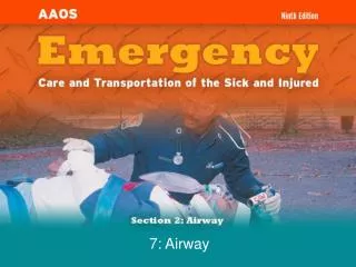
7: Airway. Cognitive Objectives (1 of 5). 2-1.1 Name and label the major structures of the respiratory system on a diagram. 2-1.2 List signs of adequate breathing. 2-1.3 List signs of inadequate breathing. 2-1.4 Describe the steps in performing the head tilt-chin lift maneuver.

E N D
Cognitive Objectives(1 of 5) 2-1.1 Name and label the major structures of the respiratory system on a diagram. 2-1.2 List signs of adequate breathing. 2-1.3 List signs of inadequate breathing. 2-1.4 Describe the steps in performing the head tilt-chin lift maneuver. 2-1.5 Relate mechanism of injury to opening the airway. 2-1.6 Describe the steps in performing the jaw-thrust maneuver.
Cognitive Objectives(2 of 5) 2-1.7 State the importance of having suction ready for immediate use when providing emergency care. 2-1.8 Describe the techniques of suctioning. 2-1.9 Describe how to artificially ventilate a patient with a pocket mask. 2-1.10 Describe the steps in performing the skill of artificially ventilating a patient with a bag-valve- mask device while using the jaw-thrust maneuver.
Cognitive Objectives(3 of 5) 2-1.11 List the parts of the bag-valve-mask system. 2-1.12 Describe the steps in performing the skill of artificially ventilating a patient with a bag-valve- mask device for one and two rescuers. 2-1.13 Describe the signs of adequate artificial ventilation using the bag-valve-mask device. 2-1.14 Describe the signs of inadequate artificial ventilation using the bag-valve-mask device. 2-1.15 Describe the steps in ventilating a patient with a flow-restricted oxygen-powered ventilation device.
Cognitive Objectives(4 of 5) 2-1.16 List the steps in performing the actions taken when providing mouth-to-mouth and mouth-to- stoma ventilation. 2-1.17 Describe how to measure and insert oropharyngeal (oral) airway. 2-1.18 Describe how to measure and insert a nasopharyngeal (nasal) airway. 2-1.19 Define the components of an oxygen delivery system. 2-1.20 Identify a nonrebreathing face mask and state the oxygen flow requirements needed for its use.
Cognitive Objectives (5 of 5) 2-1.21 Describe the indications for using a nasal cannula versus a nonrebreathing face mask. 2-1.22 Identify a nasal cannula and state the flow requirements needed for its use.
Affective Objectives 2-1.23 Explain the rationale for basic life support, artificial ventilation, and airway protective skills taking priority over most other basic life support skills. 2-1.24 Explain the rationale for providing adequate oxygenation through high inspired oxygen concentrations to patients who, in the past, may have received low concentrations.
Psychomotor Objectives (1 of 4) 2-1.25 Demonstrate the steps in performing the head tilt- chin lift maneuver. 2-1.26 Demonstrate the steps in performing the jaw- thrust maneuver. 2-1.27 Demonstrate the techniques of suctioning. 2-1.28 Demonstrate the steps in providing mouth-to- mouth artificial ventilation with body substance isolation (barrier shields). 2-1.29 Demonstrate how to use a pocket mask to artificially ventilate a patient.
Psychomotor Objectives (2 of 4) 2-1.30 Demonstrate the assembly of a bag-valve-mask unit. 2-1.31 Demonstrate the steps in performing the skill of artificially ventilating a patient with a bag-valve- mask device for one and two rescuers. 2-1.32 Demonstrate the steps in performing the skill of artificially ventilating a patient with a bag-valve- mask device while using the jaw-thrust maneuver. 2-1.33 Demonstrate artificial ventilation of a patient with a flow-restricted, oxygen-powered ventilation device.
Psychomotor Objectives (3 of 4) 2-1.34 Demonstrate how to artificially ventilate a patient with a stoma. 2-1.35 Demonstrate how to insert an oropharyngeal (oral) airway. 2-1.36 Demonstrate how to insert a nasopharyngeal (nasal) airway. 2-1.37 Demonstrate the correct operation of oxygen tanks and regulators. 2-1.38 Demonstrate the use of a nonrebreathing face mask and state the oxygen flow requirements needed for its use.
Psychomotor Objectives (4 of 4) 2-1.39 Demonstrate the use of a nasal cannula and state the flow requirements needed for its use. 2-1.40 Demonstrate how to artificially ventilate the infant and child patient. 2-1.41 Demonstrate oxygen administration for the infant and child patient.
Additional Objectives* • Describe how to perform the Sellick maneuver (cricoid pressure). • Explain the rationale for applying cricoid pressure. • Demonstrate how to perform the Sellick maneuver (cricoid pressure). • These are noncurriculum objectives.
Breathing Process: Inhalation • Active part of breathing • Diaphragm and intercostal muscles contract, allowing the lungs to expand. • The decrease in pressure allows lungs to fill with air. • Air travels to the alveoli where exchange of gases occurs.
Breathing Process: Exhalation • Does not normally require muscular effort • Diaphragm and intercostal muscles relax. • The thorax decreases in size, and ribs and muscles assume their normal positions. • The increase in pressure forces air out.
Gas Exchange • Inhalation delivers oxygen-rich air to alveoli. • Oxygen diffuses into the blood. • Breathing is primarily adjusted by the level of carbon dioxide in the blood.
Hypoxia • Not enough oxygen for metabolic needs • Develops when patient is: • Breathing inadequately • Not breathing
Signs of Hypoxia • Nervousness, irritability, and fear • Tachycardia • Mental status changes • Use of accessory muscles for breathing • Difficulty breathing, possible chest pain
Conditions Resulting in Hypoxia • Chest injury • Shock • Lung disease • Asthma • Premature birth • Myocardial infarction • Pulmonary edema • Acute narcotic overdose • Smoke inhalation • Stroke
Recognizing Adequate Breathing • Normal rate and depth • Regular pattern • Regular and equal chest rise and fall • Adequate depth
Normal Respiration Rates • Adults 12 to 20 breaths/min • Children 15 to 30 breaths/min • Infants 25 to 50 breaths/min
Recognizing Inadequate Breathing • Fast or slow rate • Irregular rhythm • Abnormal lung sounds • Reduced tidal volumes • Use of accessory muscles • Cool, damp, pale or cyanotic skin
Head Tilt–Chin Lift • Kneel beside patient’s head. • Place one hand on forehead. • Apply backward pressure. • Place tips of finger under lower jaw. • Lift chin. Head tilt-chin lift
Jaw-Thrust Maneuver • Kneel above patient’s head. • Place fingers behind angle of lower jaw. • Use thumbs to position the lower jaw.
Assessment of the Airway (1of 2)
Assessment of the Airway (2 of 2) • Assess whether breathing has returned using look, listen, and feel technique. • Listen by placing your ear about 10 inches above patient’s nose and mouth. • Feel and listen for movement of air. • Watch the patient’s chest and abdomen. • Place a hand on patient’s chest to feel for movement.
Severe Airway Obstruction • There will be no movement of air. • Chest and abdomen may rise and fall with patient’s attempts to breathe. • Chest wall movement alone does not indicate breathing. • Always use three-part approach: look, listen, and feel for movement of air.
Basic Airway Adjuncts (1 of 6) • Oropharyngeal airways • Keep the tongue from blocking the upper airway • Allow for easier suctioning of the airway • Used in conjunction with BVM device • Used on unconscious patients without a gag reflex
Basic Airway Adjuncts (2 of 6) Inserting an oropharyngeal airway 1. Select the proper size airway. 2. Open the patient’s mouth. 3. Hold the airway upside down and insert it in the patient’s mouth. 4. Rotate the airway 180° until the flange rests on the patient’s lips.
Basic Airway Adjuncts (4 of 6) • Nasopharyngeal airways • Conscious patients who cannot maintain airway • Can be used with intact gag reflex • Should not be used with head injuries or nosebleeds
Basic Airway Adjuncts (5 of 6) Inserting a nasopharyngeal airway 1. Select the proper size airway. 2. Lubricate the airway. 3. Gently push the nostril open. 4. With the bevel turned toward the septum, insert the airway.
Basic Airway Adjuncts (6 of 6) 1 2 3 4
Suction Equipment (2 of 2) French, or whistle-tip, catheter
Suctioning Technique (1 of 2) • Check the unit and turn it on. • Select and measure proper catheter to be used. • Open the patient’s mouth and insert tip. • Suction as you withdraw the catheter. • Never suction adults for more than 15 seconds.
Suctioning Technique (2 of 2) 2 1 3 4
Supplemental Oxygen • All patients in cardiac arrest should get oxygen. • Any patient with a respiratory or cardiac emergency needs oxygen. • Never withhold oxygen from anyone who may benefit from it.
Supplemental Oxygen Equipment • Oxygen cylinders • Available as a compressed combustible gas • Available in several sizes • Pin-indexing safety system • Oxygen regulators • Humidified oxygen
Oxygen Flowmeters • Pressure-compensated flowmeter • Affected by gravity; must be kept upright • Bourdon-gauge flowmeter • Not affected by gravity; can be used in any position
Using Supplemental Oxygen (1 of 2) • Inspect cylinder and markings. • “Crack” the cylinder. • Attach the regulator/flowmeter. • Open the cylinder. • Attach proper delivery device to flowmeter.
Using Supplemental Oxygen (2 of 2) • Adjust flowmeter to desired flow rate. • Apply the oxygen device to the patient. • When done, discard the delivery device. • Turn off the flowmeter.
Hazards of Oxygen • Oxygen supports combustion. • Keep possible ignition sources away from the area. • Oxygen tanks are under high pressure.
Oxygen Delivery Equipment • Nonrebreathing mask • Provides up to 90% oxygen • Used at 10 to 15 L/min • Nasal cannula • Provides 24% to 44% oxygen • Used at 1 to 6 L/min
Methods of Ventilation • Mouth to mask • Two-person BVM device • Flow-restricted, oxygen-powered device • One-person BVM device Bag-valve-mask
Rate of Artificial Ventilations Adult — 1 breath every 5-6 seconds Children — 1 breath every 3-5 seconds Infants — 1 breath every 3-5 seconds Bag-valve-mask
Mouth-to-Mask Technique (1 of 2) • Kneel at patient’s head and open airway. • Place the mask on the patient’s face. • Take a deep breath and breathe into the patient for 1 second. • Remove your mouth and watch for patient’s chest to fall.