Download

1 / 21
510 likes | 1.39k Views
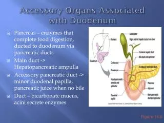
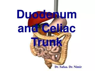
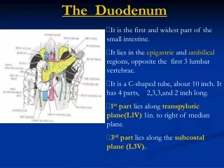
Duodenum. It is the shortest, widest and most fixed part of the small intestine. Position and Shape: Fixed to the posterior abdominal wall (retroperitoneal), occupies the epigastric and umbilical regions. Follows a C- shaped course around the head of the pancreas.

E N D
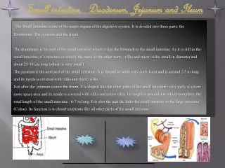
It is the shortest, widest and most fixed part of the small intestine. • Position and Shape: • Fixed to the posterior abdominal wall (retroperitoneal), occupies the epigastric and umbilical regions. • Follows a C- shaped course around the head of the pancreas. • Extends from the pylorus to the duodeno-jejunal flexure.
Parts of the duodenum: • 1. First part: - 2 inches long, its 1st inch is mobile as it is covered by peritoneum ant. and post. - It begins at the pylorus, 1/2 an inch to the right of the median plane at the level of L1 (transpyloric plane).
Relations of 1st part: • Anteriorly: quadrate lobe of the liver and gallbladder. • Posteriorly: - Neck of the pancreas, - Portal vein, - bile duct
Inferiorly: head of pancreas. • Superiorly: related to epiploic foramen.
2. Second part: -3 inches long and descends vertically from the level of L1 to L3. Covered by peritoneum anter. • Relations: - Anterior: right lobe of the liver, gall bladder, transverse colon and coils of jejunum. - Posterior: hilum of the right kidney. - Lateral: hepatic flexure of the large intestine. - Medial:head of pancreas, bile duct and pancreatico –duodenal arteries.
3. Third part: 4 inch long, lies horizontally opposite to the level of L3. • Relations: - Anterior:superior mesenteric vessels and coils of small intestine. - Posterior:it is related to the following structures from the right to the left side:right ureter, right psoas major, right. gonadal vessels, IVC, aorta and inferior mesenteric artery. - Superior: head of pancreas. - Inferior: coils of small intestine.
4. Fourth part: - It is one inch long. It ascends from the level of L3 to L2 one inch to the left of median plane at the dudeno-jejunal flexure.
Relations of the 4th part of theduodenum: • Anterior:transverse colon and transverse mesocolon.
Posterior:left psoas major, left gonadal and left renal vessels. • Medial:head of pancreas and aorta. • Lateral: Lt Kidney.
Arterial supply of the duodenum: • 1. Supra-duodenal artery: from the hepatic artery proper (coeliac trunk). • 2. Superior pancreatico-duodenal artery: from gastro-duodenal (coeliac). • 3. Inferior pancreatico-duodenal artery: from superior mesenteric artery.
Peritoneal recesses of the duodenum: - Superior duodenal recess. - Inferior recess. - Paraduodenal recess. - Retroduodenal recess.
COELIAC TRUNK • Origin: front of abdominal aorta ,upper border of L1. • Course: short trunk passes horizontally between two crura of diaphragm.
Regions supplied by it: it supplies the foregut. • Branches: Main branches are: • Left gastric artery • Hepatic artery • Splenic artery
Branches of coeliac trunk • Left gastric artery: • Course: upward, to left till diaphragm ,curves down along lesser curvature ,between layers of lesser omentum ,then anastomoses with Rt gastric artery.
Branches: • Oesophageal branches. • Gastric branches.
2- Hepatic artery • Branches: • Right gastric artery. • Supra-duodenal artery. • Gastroduodenal artery. • Right gastroepiploic artery. • Superior pancreaticoduodenal artery. • Right hepatic artery (cystic a.) • Left hepatic artery.
3- Splenic artery • it passes transversely to the left behind stomach. • Along upper border of pancreas. - It has a tortuous course. • It’s accompanied by splenic vein. • It passes In front of left suprarenal gland and left kidney where it passes between two layers of lienorenal ligament to reach the hilum of spleen.
Branches: 1- Pancreatic branches: supply pancreas 2- Short gastric arteries: to upper part of greater curvature of the stomach. 3- Left gastroepiploic artery:it passes between two layers of greater omentum along the greater curvature of stomach .
Termination: It terminates by dividing into 5 or 6 splenic arteries which enter the hilum of spleen .
Thank You Prof.: Dr. Shawky Tayel