Download
1 / 28
290 likes | 529 Views
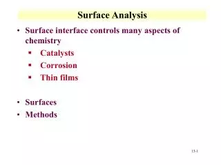
Protein Surface Analysis for Functional Analysis and Prediction. T. Andrew Binkowski and Andrzej Joachimiak 2009 NIGMS Workshop: Enabling Technologies for Structural Biology March 4-6, 2009. Outline. How Can Surface Analysis Aid Your Structural Genomics Effort? Protein Surfaces

E N D
Protein Surface Analysis for Functional Analysis and Prediction T. Andrew Binkowski and Andrzej Joachimiak 2009 NIGMS Workshop: Enabling Technologies for Structural Biology March 4-6, 2009
Outline How Can Surface Analysis Aid Your Structural Genomics Effort? • Protein Surfaces • Comparing Surfaces of Proteins • Surface Analysis in the Structural Genomics Pipeline • The Global Protein Surface Survey
Functional Inference in Proteins • Transfer function based on similarity to a protein with known biological activity • Sequence • 30-70% • Functional sites result from spatial interactions of key residues in diverse regions of primary sequence • Structure • Reveal more distant relationships • 1 fold ~ many functions; vice versa • Example: generalized secondary structural element • Different SSE can bring residues in spatial proximity • (Jaroszewski & Godzick, ISMB 00)
Functional Inference in Proteins • Functional surfaces may be the most conserved structural features of proteins • Surfaces performing identical biochemical activity can be found within different protein scaffolds or in the absence clear evolutionary relationships • Exploit ability of proteins to preserve local spatial residue patterns • Presents another opportunity to infer insightful ideas about their biological function and mechanisms • Novel heme-monooxygenase • 12% sequence identity • a/b vs. all a • Experimentally verified activity
Surfaces of Proteins • Surface: • Local grouping of solvent accessible atoms • Pockets: • Empty concavity on a protein surfaces into which solvent can gain access • Identifying surfaces: • Methods: • Solvent accessibility, Geometry, Grids, Spheres • Applications: • CASTp, Surfnet, Pocket, Ligsite, Pass • Our approach: • Computational geometry (alpha shape) • CASTp, PDB, Swiss-Prot, Catalytic Site Atlas • Ligand binding surfaces: • Exclusion contact surface (solvent accessibility difference) • Muck & Edelsbrunner, ACM Tran Graph, 1994; Edelsbrunner, Facello, Liang, Disc Appl Math, 1996; Liang, Edelsbrunner, Woodward, Protein Sci, 1998
Global Protein Surface Survey http://gpss.mcsg.anl.gov
SurfaceScreen Methodology for identifying similarly shaped proteins and aligning them Optimizes two components Global Shape Perceived similarity Size and scale, independent of chemistry Local physicochemical texture Preserved atom/residue orientation Conservation of chemical complimentarity Comparing Surfaces of Proteins Surface Global Surface Shape Filtering Surface Shape Alignment Constrained Spatial Surface Refinement Apply Scoring Functions
Comparing Surfaces of Proteins:Global Shape Similarity • Surface Shape Signatures (SSS) • Represent signature of a surface as distribution sampled from a shape function (Osada et. Al., 2002) • Comparison of probability distributions • Kolmogorov-Smirnov • Earth Mover’s Distance • ATP Binding sites • protein kinase CK2 from Z. mays (b) • phosphopantetheine adenylyltransferase from E. coli (c) • maltose/maltodextrin transport protein from E. coli (d,cyan chain A, light blue chain B) • 50 non-homologous sites (< 30% sequence identity)
Spatial Surface Alignment Refinement • Combinatorial comparison of residue sets in “neighborhood” • Maintain “like” correspondence of types • Maximum common residues • Enumerate and evaluate alignment orientations • Find optimal superposition using SVD of correlation matrix (Umeyama 1991) • Heme binding pockets of myoglobin from different organisms.
Evaluating Surface Alignments • Surface Volume Overlap: • Interpretation of SVOT is not straightforward • Need global and local • RMSD Distance: • Estimate the probability of obtaining a specific RMSD for nres • Compute random surface alignments (108) and build lookup tables • RMSD variants: • cRMSD (coordinate) • oRMSD (orientation)
Heme Binding Site Retrieval • Heme (iron-protoporphyrin IX) • Multi-functional (i.e.oxygen binding/transport, electron transfer and redox) • Binding on 20 different folds • Between proteins <2% seq. id. • Query myoglobin (gray) against PDB structure to identify hemoproteins • Retrieval rate (area under ROC curve) • Sequence: 68.7% • Structure (SSM): 64.4% • Surface: 95.8% • Detection of convergent heme binding site on IsdG from S. aureus • Missing characteristic sequence motif • 12% seq id; different scaffold • Experimentally verified monooxygenase activity • seq. & fold • surface analysis
ATP: Retrieval of a Flexible Ligand • Adenosine 5’-triphosphate multifunctional nucleotide (i.e.cell signaling, enegry transfer) • 58 unique EC classifications #.#.#.# • Conformational flexibility • Retrieval rates for 4 conformations (79.1%-85.4%); method is tolerant to flexible ligands
Prediction and Validation of GDP Binding Surface • Structure of F420-0:gamma-glutamyl ligase from A. fulgidus • Large binding surface was searched to support functional predictions and GDP binding surface is identified • Posed GDP based on superposition of surfaces (red) • Co-crystallization experiments validates prediction
Exploiting Protein Surfaces in Structural Genomics • Developing surface-based tools to address specific needs of structural genomics pipeline • Ligands for co-crystallization • Aid in the assignment of electron density • Functional annotation tools • Drive further studies (i.e. ligand binding, discovery) Ligand Identification Functional Analysis Co-crystallization Mutation Future Studies Discovery
Crystallization/Structure Improvement Partially Solved or Low Quality Structure Surface Identification Search GPSS for Binding Sites Co-crystallization Experiments • Introduction of GDP to F420-0:gamma-glutamyl ligase from A. fulgidus • improves resolution from 2.8 to 1.9 Angstroms and orders loop regions.
Assisted Electron Density Assignment • Unidentified ligand density • Construct surface surrounding density and search against ligand surface library • Does not require entire structure to be built
Assisted Electron Density Assignment • Applicable to ligands of various molecular weights and sizes • Fructose (pdb id=1zx5) • NADP (pdb id=2ag8) • Suggest a list in cases of ambiguity
Landscape Analysis: ATP • Classification based on surface similarity shows functional families have preferred (not necessarily unique) surfaces and conformation
Automated Protein Kinase Classification • All-against-all surface comparison of all protein kinases in the PDB • Color labeled by expert annotation (KinBase) • Surface clustering identifies: • Dual substrate specificity of CK2 proteins • Active/inactive states • Similarity detected between MAP p38 kinase and Abelson leukemia virus tyrosine kinase (Abl) with bound cancer drug STI-571 • MAP kinase has unique DFG “out” conformation not previously seen in ser/thr kinases
Function Sleuth • Conserved protein of unknown function (VCA0319) from V. cholerae • apc29617 • Unique arrangement of common structural motifs • Problematic for secondary structure and fold analysis • Surface analysis identifies DNA binding surface and 5 putative metal binding sites • All 5 metal binding sites showed strong preference for Mg • Putative metalloregulated repressor with Mg-regulated mechanism of DNA binding
Function Sleuth 1bdb NAD 1hoh MGD 2qwr ANP 1jbw ACQ Target APC7761 (3fd3) Agrobacterium tumefaciens str. C58
Function Sleuth Target APC61725 (3fz5) Rhodobacter sphaeroides 2.4.1 • Top 17 most similar surfaces bind B12 1i9c
Global Protein Surface Survey • SurfaceScreen for PSI ‘function sleuth’ targets • Automated analysis of largest 5 surfaces (per chain and unit) • Technical Note: • DOE INCITE on Blue/GeneP at ANL http://gpss.mcsg.anl.gov
Conclusion • Comparing surfaces of proteins can be a useful tool with many applications • Functional characterization • Assisted electron density assignment • Automated classification • Global Protein Surface Survey • http://gpss.mcsg.anl.gov
Acknowledgements ANL/MCSG H. An, G. Babnigg, L. Bigelow, A. Binkowski, C-s. Chang, S. Clancy, G. Cobb, M. Cuff, M. Donnelly, C. Giometti, W. Eschenfeldt, Y. Fan, C. Hatzos, R. Hendricks G. Joachimiak, H. Li, L. Keigher, Y-c. Kim, N. Maltseva, E. Marland, S. Moy, R. Mulligan, B. Nocek, J. Osipiuk, M. Schiffer, Univ. College London @ EBI, J. Thornton, C. Orengo, M. Bashton, R. Laskowski, D. Lee, R. Marsden, D. McKenzie, A. Todd, J. Watson Northwestern Univ. W. Anderson, O. Kiryukhina D. Miller, G. Minasov, L. Shuvalova, X. Yang, Y. Tang Univ. of Toronto A. Edwards, C. Arrowsmith, A. Savchenko, E. Evdokimova, J. Guthrie, A. Khachatryan, M. Kudrytska, T. Skarina, X. (Linda) Xu ANL/MCSG A. Sather, G. Shackelford, L. Stols, K. Tan, C. Tesar, R-y. Wu, L. Volkart, R-g. Zhang, M. Zhou, ANL/SBC N. Duke, S. Ginell, F. Rotella Washington Univ. D. Fremont, T. Brett, C. Nelson, Univ. of Chicago O. Schneewind, D. Missiakas, P. Gornicki, S. Koide, ITCSG W-j. Tang, B. Roux, J. L. Robertson M.R. Rosner, T. Kossiakoff, ITCSG V. Tereshko, G. Montelione, Ruthgers Univ. NESGC T. Terwilliger, Los Alamos, ITCSG Z. Derewenda, Univ. of Virginia, ITCSG Z. Dauter, NCI J. Liang, Univ. of Illinois D. Sherman, U. Michigan Univ. of Virginia W. Minor, M. Chruszcz, M. Cyborowski, M. Grabowski, P. Lasota, P. Miles, M. Zimmerman, H. Zheng Univ. of Texas SWMC Z. Otwinowski, D. Borek, A. Kudlicki, A. Q. Mei, M. Rowicka Funding: NIH and DOE 27 27