Download
1 / 47
630 likes | 1.36k Views
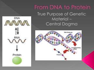
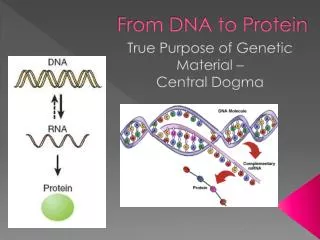
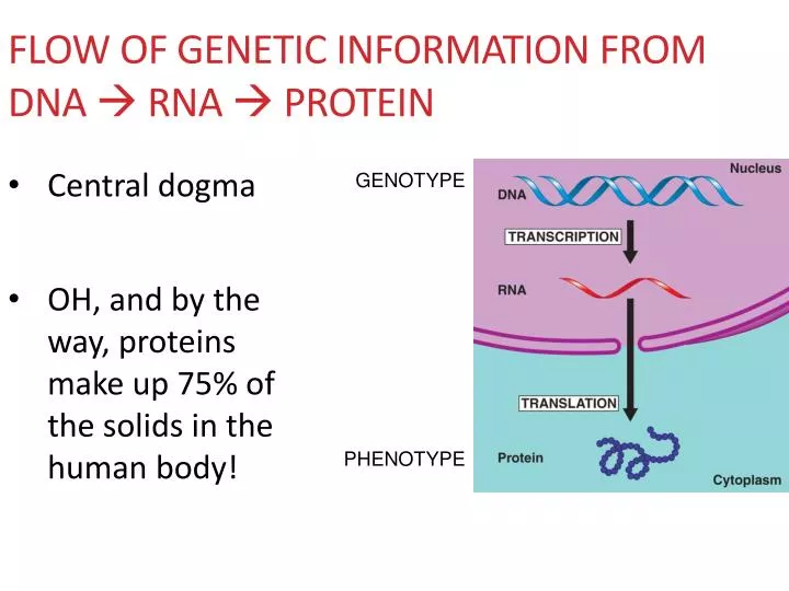
Flow of Genetic Information from DNA RNA Protein. Central dogma OH, and by the way, proteins make up 75% of the solids in the human body!. GENOTYPE. PHENOTYPE. DNA specifies synthesis of proteins in 2 stages: Transcription - the transfer of genetic info from DNA RNA molecule

E N D
Flow of Genetic Information from DNA RNA Protein • Central dogma • OH, and by the way, proteins make up 75% of the solids in the human body! GENOTYPE PHENOTYPE
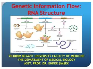
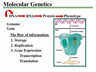
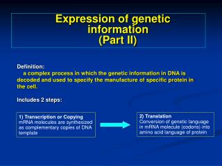
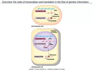
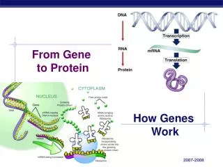
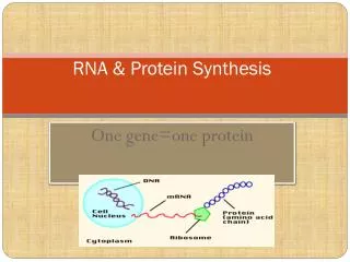
DNA specifies synthesis of proteins in 2 stages: • Transcription - the transfer of genetic info from DNA RNA molecule • Translation - the transfer of info from RNA protein
The Gene • Unit of heredity with a specific nucleotide sequence that occupies a specific location on a chromosome • E.g. Map of human chromosome 17 showing a breast cancer gene (BRCA-1) • Humans have two copies of BRCA-1 which normally suppresses breast cancer • If one copy is defective, then no back up if other gene damaged by exposure to environmental carcinogens • Inheriting a defective BRCA-1 gene risk of breast cancer
The Language of Nucleic Acids • For DNA, the alphabet is the linear sequence of nucleotide bases • A single DNA molecule may contain 1000’s of genes • A typical gene consists of 1000’s of nucleotides
http://en.wikipedia.org/wiki/File:Genome_Sizes.png Relative Genome Sizes
Transcription of DNA • DNA’s nucleotide sequence “rewritten” into RNA nucleotide sequence (remember that both are nucleic acids) • RNA is made from the DNA template, using a process resembling DNA replication except • T’s are substituted by U’s • RNA nucleotides are linked by RNA polymerase
Unpacking Transcription • Three phases • Initiation • RNA elongation • Termination
Initiation of Transcription • “Start transcribing” signal is nucleotide sequence, called a promoter • Located at beginning of gene • RNA polymerase attaches to the promoter (via transcription factor) • RNA synthesis begins
RNA Elongation • RNA grows longer • RNA strand peels away from the DNA template
Termination of Transcription • RNA polymerase reaches specific nucleotide sequence, called a terminator • Polymerase detaches from RNA • DNA strands rejoin
Processing of Eukaryotic RNA • Unlike prokaryotes, eukaryotes process their RNA • Add a cap & tail - xtra nucleotides at ends of RNA transcript for protection (against cellular enzymes) & recognition (by ribosomes later on) • Removing introns – stretches of noncoding nucleotides that interrupt coding stretches = the exons • Splicing exons together to form messenger RNA (mRNA)
Translation • Conversion from nucleic acid language to protein language • Requires • mRNA • ATP • Enzymes • Ribosomes • Transfer RNA (tRNA)
The Genetic Code • Shared by ALL organisms • The set of rules that relates mRNA nucleotide sequence to amino acid sequence • Since there are 4 nucleotides, there are 64 (or 43) possible nucleotide “triplets” = codons • 61 codons code for amino acids, 3 act as “start” or “stop” codons marking the beginning or end of a polypeptide http://www.nature.com/scitable
The Genetic Code Fig. 10.11
tRNA • Acts as molecular interpreter – decodes mRNA codons into a protein • Each codon (thus amino acid) is recognized by a specific tRNA • Has an anticodon – recognizes & decodes an mRNA codon • Has amino acid attachment site • When tRNA recognizes & binds • to its corresponding codon in • ribosome, tRNA transfers its • amino acid to the end of the • growing amino acid chain
Ribosomes • Organelles that • coordinate functions of mRNA & tRNA during translation • contain ribosomal RNA (rRNA)
Unpacking Translation • Occurs in the ribosome • Like transcription, broken down into 3 phases • Initiation • Elongation • Termination • Short but sweet translation animation • http://www.nature.com/scitable/content/translation-animation-6912064
Initiation of Translation • Small ribosomal subunit binds to start of the mRNA sequence • Then, initiator tRNA carrying the amino acid methionine binds to the start codon of mRNA • Start codons in all mRNA molecules are AUG and code for methionine! • Next, large ribosomal subunit binds
Polypeptide Elongation • Large ribosomal unit binds each successive tRNA w/ its attached amino acid • Ribosome continues to translate each codon • Each corresponding amino acid is added to growing chain and linked via peptide bonds • Elongation continues until all codons are read.
Termination of Translation • Occurs when ribosome reaches stop codon (UAA, UAG, & UGA) • No tRNAmolecules can recognize these codons, so ribosome recognizes that translation is complete. • New protein released • Translation complex dismantles • into its subunits
Termination of Translation • sdf Fig. 10.20
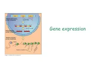
Transcription & translation are how genes control • structures • activities of cells • In other words, FORM & FUNCTION of proteins!
DAY 5: cell Structure & function IMSS BIOLOGY ~SUMMER 2011
Major Categories of Cells Prokaryotic cells (the prokaryotes) – vast spp diversity & abundance !!! Domain Archaea - all Domain Bacteria -all Eukaryotic cells (the eukaryotes) Domain Eukarya - mostly
Genetic Diversity • Microbes make up most of Earth’s genetic diversity • This “tree of life” is like a map of genetic relatedness • Distance (line length) genetic relatedness Norm Pace, U. Colorado
Three-domain Classification System Bacteria & Archaea diverged very early in evolutionary history Archaea more closely related to Eukarya
Extremophiles & the Search for Life Beyond Earth We’ve found prokaryotes in virtually EVERY place on Earth, even the most unlikely (extreme) places Extremophiles: organisms that live in “extreme” environments Scientists are studying these microbes for a better idea of life’s capacities AND the potential of extra-terrestrial life
NASA and Microbes • Microbes @ NASA • Loads of research, e.g. • Extremophiles • How life evolved on Earth • Biomedical applications • Modes of virulence & pathogenesis
Mono Lake Bacteria: Recent Discovery Oremland & Kulp, USGS, Science (2008) https://www.sciencemag.org/cgi/content/abstract/321/5891/967 First e.g. of photoautotroph that also uses arsenic to “fix” CO2 Microbial arsenic metabolism may extend back to primordial Earth
Rio Tinto, Spain 5,000 yrs. of mining activity Extreme acidity Extreme heavy metal concentrations Surprisingly more eukaryote than prokaryote diversity “On Earth, microbial communities thrive in highly acidic waters rich in iron and sulfur, such as the blood-red waters of the Rio Tinto in southwestern Spain. Among the minerals dissolved in the Rio Tinto is jarosite, an iron- and sulfur-bearing mineral also found on Mars.” -- http://amesnews.arc.nasa.gov/releases/2003/03_74AR.html
A Bacterial Superhero • Deionococcusradiodurans • Found to “beat the constraints” for survival on Mars (R. Richmond et al., NASA’s Marshall Space Flight Center) • Radiation • Cold • Vacuum • Oxidative damage
Core Principle The cell Basic unit of life Multicellular organisms are organized structures made up of different cells Ea. cell shares common properties w/ other cells Ea. cell has some specialized structures & functions Cell size (& function) is limited by surface area (SA) to volume (V) relationships
ACTIVITY • SA/V Relationship – Tory Brady min.
Which cell shape would be best in places where rapid exchange of substances (via diffusion) is a high priority? C B A
SA/V Ratios • Can be applied to • single cells (including single-celled organisms) • Important when considering transport mechanisms and cell size limitations • whole animals • Important when considering metabolic and thermoregulatory principles
Small intestine (SI) Histology Form follows function: SI microanatomy important to understanding its function SI completes digestion of food, and most of all nutrient absorption occurs here !!!
Structure of intestinal mucosa allows for a 600x greater luminal surface area than if it had a flat surface Intestinal folds 3x in SA Villi 10x in SA Microvilli 20x in SA
The Scale of Life • How can we “see” the tiniest organisms (or their components)? • The unaided human eye is limited to ~0.1 mm • How can we see things smaller than this? . m m m
We need to use microscopy to magnify & resolve very tiny objects to > 1 mm in order to “see” them http://www.cellsalive.com/howbig.htm
Key Factors of Microscopy • Magnification • How much larger object appears w/ microscope lenses than w/out • Resolution • Amount of detail (ability to distinguish between 2 pts. on an image) http://homepages.gac.edu/~cellab/chpts/chpt1/intro1.html
Microscopy - Overview • Many types for different levels of detail
Light Microscopes • Most widely used & available • Basic anatomy • Total magnification = eyepiece lens power x objective lens power • http://www.under-microscope.com/
Microscopy - Resources • Thorough coverage of the various types of microscopy, how they work, & their functions • http://www.cas.muohio.edu/~meicenrd/ANATOMY/Ch1_Microscopy/microscopy.html • More basic descriptions of microscope types along with an excellent photo/video library • http://www.under-microscope.com/
ACTIVITIES • Cells alive – Termite Guts – Tory Brady • Tools of the trade – microscopy • Digital microscopy in the classroom – Sandi Yellenberg 60 min.