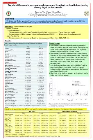
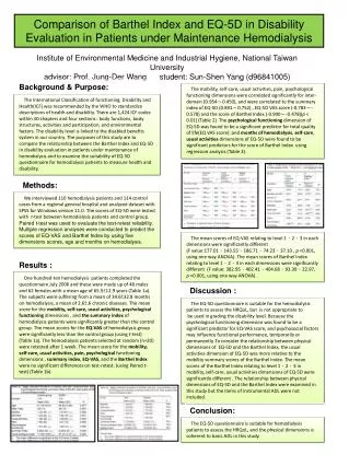
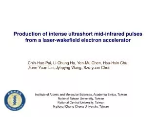
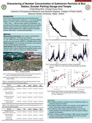
Download
1 / 18
180 likes | 329 Views
The proteomics approach to study the role of Helicobacter pyroli in the development of gastric cancer. Lu-Ping Chow Graduate Institute of Biochemistry and Molecular Biology National Taiwan University.

E N D
The proteomics approach to study the role of Helicobacter pyroli in the development of gastric cancer Lu-Ping Chow Graduate Institute of Biochemistry and Molecular Biology National Taiwan University
World distribution of H. pylori infection and its gastric consequences from common chronic gastritis
Class I carcinogen of GC [WHO, 1994] GC vs. H. pylori • The prevalence of H. pylori in GC patients is much higher than in age- and gender-matched controls. • The association between H. pylori positivity on serology and overall gastric cancer risk is higher than 60% . H. p+ GC Scand J Gastroenterol 37:891–898.(2002)
Importance steps during H. pyroli interaction with its host in the gastric mucosa
Host responses induced by H. pylori : 1. Inflammatory response Med Sci Monit9:sr 53-66 (2003)
Host responses induced by H. pylori : 2. Proliferation (G1/S transition) JOURNAL OF CELLULAR PHYSIOLOGY200:334–342 (2004)
Host responses induced by H. pylori : 3. Induction or prevention gastric epithelial-cell apoptosis NATURE REVIEWS CANCER 2:28-37(2002)
Sub-cellular and functional proteomic analysis of the cellular responses induced by Helicobacter pylori AGS cells AGS cells co-cultured with H. pylori Histogram View Image View 3D view Table View 1000 1500 2000 Mass (m/z) Sample #1 Protein Name Index # Acession # Score Protein 1 Probable DNA-Directed RNA Polym 32443 P05472 0.854 Protein 2 Mitochondrial Respiratory Chain 30371 P40341 0.731 Protein 3 Tyrosine Protein Kinase SRC64B 34968 P00528 0.921 Strategy Cy5-labeled non-infected cells Cell lysis and fractionation CyDye labeling & 2D gel seperation Cy2-labeled pooled standard Cy3-labeled infected cells Excision of spots In-gel digestion Elution of fragments Database search Quantification of Decyder Identification targets by peptide mass fingerprinting (PMF)
Functional analyses of effectiveness of H. pylori infection of AGS cells using a MOI of 100. Scale bar =10μm. Fig.1 • Induction of the scattering (" hummingbird" ) phenotype of AGS cells after infection with H. pylori for 4 h. • IL-8 release of AGS cells after infection with H. pylori for 24 h by ELISA. • Induction of COX-2 protein expression in AGS cells after infection with H. pylori for 24 h by immunoblot analysis.
Sample loading : 50ug per lane HGFR : indicator of membrane fraction Urease A : indicator of H. pylori contaminant Cell fractionaion of non-infected and H. pylori-infected AGS cells Fig.2 UreaseA was present at high amounts in the bacteria plus cell pellet fraction of H. pylori-infected cells, present at low amounts in the membrane fraction, and absent in the host cytosol fraction.
C Dye swapping strategy was adopted to avoid dye labeling-bias, therefore, Cy3 and Cy5 dyes were interchangeable. 2D DIGE analysis of alterations in the cytosolic fraction of AGS cells induced by H. pylori infection A • H. pylori-infected : green non-infected : red Fig.3
TABLE I Proteins in the cytosolic fraction of AGS cells showing up- or down-regulation after 24 h of H. pylori infection identified by MS
cell communication and signal transduction proteins cytoskeleton proteins others transcription and translation-related proteins 3.6% 7.1% 7.1% 25% oxygen-regulated proteins 7.1% 21.4% 7.1% 17.9% protein synthesis and folding-related proteins angiogenesis/ metastasis-related proteins metabolic enzymes Bioinformatics ontology of the identified proteins • 2-fold up-regulation after H. pylori-infection • Potential cancer-associated proteins
2-DE immunoblot analysis and three-dimensional fluorescence intensity profiles of non-infected AGS cells and AGS cells infected with H. pylori Fig.4 The greatest changes were seen for laminin γ-1, VCP, HSP70, and 14-3-3 β, while moderate changes were seen for FKBP4, MMP-P1, TCP1α, and enolase α.
Immunoblot analysis of expression profiles of lamininγ-1, VCP, HSP70, TCP 1, MMP-P1, FKBP4, Enolaseα, and 14-3-3βin paired cancerous (T) and noncancerous (N) gastric tissues Fig.5 Increased spots were seen in 9 of the 10 paired samples for laminin γ-1, 6 for VCP, 7 for HSP70, 7 for MMP-P1, 10 for FKBP4, 6 for TCP1, 10 for enolase α, and 10 for 14-3-3 β.
Immunohistochemical study of VCP, TCP 1, MMP-P1, Enolase, and 14-3-3βin gastric cancer tissue Fig.6 Expression of VCP, MMP-P1, TCP1, enolaseα, and 14-3-3βwas more abundance in gastric canceorus cells than in paired normal cells whereas most cases had similar expression amount of lamininγ-1, HSP70 and FKBP4 proteins.
Summary 1. An in vitro model was established using a MOI 100 and evaluating the effectiveness of H. pylori infection by functional analyses. 2. Twenty-seven differential expressed proteins in H. pylori- infected AGS cells were identified by proteomic approach. 3. The identified protein were classified as cytoskeleton proteins, protein synthesis and folding-related proteins, metabolic enzymes, angiogenesis/metastasis-related proteins, oxygen- regulated proteins, transcription and translation-related proteins, or cell communication / signal transduction-related proteins by bioinformatics ontology. 4. Valosin-containing protein, mitochondrial matrix protein P1, T-complex protein 1, enolaseα and 14-3-3βwere found to be overexpressed in cancerous tissues by immunoblot assay and immunohistochemical staining.