Download
1 / 44
480 likes | 658 Views
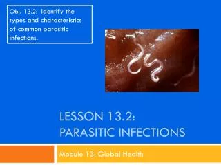
Lecture 7 clinical practice PARASITIC INFECTIONS BASICS IN DIAGNOSIS. By dr. Dalia Galal. BASIC TERMINOLOGY AND PRINCIPLES Symbiosis: Living together Commensalism: One symbiont benefits, other unaffected Mutualism: Both symbionts benefit

E N D
Lecture 7 clinical practicePARASITIC INFECTIONS BASICS IN DIAGNOSIS By dr. Dalia Galal
BASIC TERMINOLOGY AND PRINCIPLES • Symbiosis: Living together • Commensalism: One symbiont benefits, other unaffected • Mutualism: Both symbionts benefit • Parasitism: One symbiont benefits, other is damaged
Key definitions: What is ….? • Medical parasitology: “the study and medical implications of parasites that infect humans” • A parasite: “a living organism that acquires some of its basic nutritional requirements through its contact with another living organism”. Parasites may be simple unicellular protozoa or complex multicellular metazoa • Eukaryote: a cell with a well-defined chromosome in a membrane-bound nucleus. All parasitic organisms are eukaryotes 3
Key definitions: What is ….? • Protozoa: unicellular organisms, e.g. Plasmodium (malaria) • Metazoa: multicellular organisms, e.g. helminths (worms) and arthropods (ticks, lice) • An endoparasite: “a parasite that lives within another living organism” – e.g. malaria, Giardia • An ectoparasite: “a parasite that lives on the external surface of another living organism” – e.g. lice, ticks 4
Key definitions: What is ….? • Host: “the organism in, or on, which the parasite lives and causes harm” • Definitive host: “the organism in which the adult or sexually mature stage of the parasite lives” • Intermediate host: “the organism in which the parasite lives during a period of its development only” • Zoonosis: “a parasitic disease in which an animal is normally the host - but which also infects man” • Vector: “a living carrier (e.g.an arthropod) that transports a pathogenic organism from an infected to a non-infected host”. A typical example is the female Anopheles mosquito that transmits malaria 5
Laboratory Methods For Parasites In Faeces • If technique is 100% successful in detecting parasites by a single stool examination, at least three serial stools must be examined before a patient can be considered free from infections in which stages of parasites would be expected to be found in the faeces. • Whilst clinical symptoms or a case history may provide clues as to which parasites may be present, each faecal specimen should be treated as an unknown, as parasite stages unrelated to the clinical picture may be present.
Faecal specimens may contain several stages of Parasites • Faecal specimens are examined for the presence of protozoa and helminthes larvae or eggs. • The stages of protozoa found in stools are trophozoites and cysts. • The stages of helminthes usually found in stools are eggs and larvae, though whole adult’s worms or segments of worms may also be seen. • Adult worms and segments of tapeworms are usually visible to the naked eye, but eggs, larvae, trophozoites, and cysts can be seen only with the microscope.
Collection of faecal specimens • Because of the fragile nature of many intestinal parasites, and the need to maintain their morphology for accurate identification, reliable microscopic diagnosis can’t be made unless the stool is collected properly. • Approximately 10 grams of fresh faeces uncontaminated by urine, into a clean plastic container. 3. The container should be free from antiseptics and disinfectants. 4. Label all samples clearly with the patient’s name, reference number, date, and time of collection.
Collection of faecal specimens • All samples should be accompanied by a requisition form from the physician giving relevant clinical details and recent travel history. 6. Samples from patients with a confirmed or suspected diagnosis of certain infectious diseases such as AIDS or hepatitis should be clearly labeled with “Risk of Infection”.
Collection of faecal specimens 7. Most viable parasites are susceptible to desiccation or temperature variation. If time lapse between collection and observation is considerable, i.e. more than 4 days, it may be necessary to add some form of preservative to the faeces to retain the morphology as near to the original as possible. 8. Formed samples can be kept in a refrigerator at – 4oc for a short while, but not in incubator.
Collect the Information of the Patient • History (Age, occupation, residency, previous infection) • Complaint • Clinical examination • Investigations - Laboratory investigations - Radiology - Surgical Provisional diagnosis Confirm the diagnosis
The Microscopy in Parasitology • The Microscope is the parasitologist’s main tool. Most suitable objectives are the x10, x40, and x100. • The Microscope must be covered and immersion oil removed from the lens -with xylene or ether when not in use. • Calibration of the Microscope Eyepiece Micrometer: • On many occasions measuring different sizes of suspected parasites in faeces is helpful for identification. (eyepiece micrometer)
Microscopic Examination of Wet Mount • Wet mount is the simplest and easiest technique for the examination of faeces, and this method should be performed in all laboratories. • A wet mount can be prepared directly from faecal material or from concentrated specimens. The basic types of wet mount that should be used for each faecal examination are saline, iodine, and buffered methylene blue.
The saline wet mount • Is used for the initial microscopic examination of stools. • It is employed primarily to demonstrate worm's eggs, larvae, protozoan trophozoites, and cysts. • This type of mount can also reveal the presence of red blood cells and white blood cells.
The Iodine wet mount • Is used mainly to stain glycogen and the nuclei of cysts, if present. • Cysts can usually be specifically identified in this mount. The buffered methylene blue (BMB) wet mount should be prepared each time amoebic trophozoites are seen in a saline wet mount, or when their presence is suspected.
Direct saline and iodine mounts 1. With a wax pencil writes the patient’s name or number and the date at the left-hand end of the slide.
Preparing a faecal specimen • Place a drop of saline in the centre of the left half of the slide and place a drop of iodine solution in the centre of the right half of the slide. • Note: If the presence of amoebic trophozoites is suspected, warm saline (37c) should be used.
Preparation of Wet film • With an applicator stick (match or tooth pick), pick up a small portion of the specimen (size of a match head) and mix the drop of saline.
Lecture 8 clinical practice PARASITIC INFECTIONS (II) By dr. Dalia Galal
Saline smear Iodine smear STOOL EXAMINATIONA Rapid Methods saline Iodine 1% • Huge number of: • Eggs • Protozoal troph. Motility • (Amoeb, flagellates) • Huge number of: • Cyst morphological details
Need for Concentration Methods for Faecal examination • A concentration procedure is performed mainly to separate the parasites from faecal debris. The concentration procedure not only increases the numbers of parasites in the sediment but it also unmasks them, making them more visible by removing organic and inorganic debris
STOOL EXAMINATIONScanty infectionConcentration techniques Sedimentation Floatation The sedimentation technique is a qualitative method for detecting trematode eggs in the faeces. Most trematode eggs are relatively large and heavy compared to nematode eggs It is based on the separating of eggs from faecal material and concentrating them by means of a flotation fluid with an appropriate specific gravity • Non Operculated eggs • Cestode • Nematode(Hookworms,Trichostong) • Cysts • Heavy eggs (Ascaris egg) • Operculated eggs (Trematodes) • Larvae • Cysts
STOOL EXAMINATIONSaline sedimentation Mesh wire gauze Saline Emulsify Conical flask 10 g stool Sediment
Kato techniqueit involves staining a sieved fecal sample and examining it under a microscope. The total number of stained eggs are counted and used to calculate the number of eggs per gram Mesh screen Hole Template Remove the template Cellophane soaked by glycerin (clears faeces( Egg count/ g stool Egg quant. Of: Ascaris, Trichomonasvaginalis, Hookworms, Schistosomamansoni
Stool examination other Techniques • Stoll’s technique for eggs of Ascaris, T. trichiura., Hookworms, S. mansoni. The Stoll Egg Counting Technique is a method for determining the number of nematode eggs per gram of feces in order to estimate the worm load in host. The advantage of this technique is that it requires no specialized equipment. The disadvantage is that the counting takes a long time because of the amount of extra (non-egg) material on the slides. • Baermann’s technique Detection.The Baermann technique is used to isolate lungworm larvae from faecal samples and infective larvae from faecal cultures. It is based on the active migration of larvae from faeces suspended in water and their subsequent collection and identification. • Cultures for Nematode larvae using Filter paper culture for Larvae of: Hookworms
STOOL EXAMINATION Stoll’s technique Egg quant. Of: Ascaris, Hookworms, S. mansoni 24 hr stool 60 CC 4 g Stool 56 CC NaOH Shake well 0.15 CC Egg count/ slide Eggs/1g= Eggs/slideX100 Erlynmeyer flask Egg/day=Eggs/1g X stool wt/g in 24 hrs
STOOL EXAMINATION Baermann’s technique Stool/soil seive 25-50CC Warmwater Glass funnel 30 min • centrifuge clamp Detec. Of Nematode L. /stool, soil
STOOL EXAMINATION Cultures for Nematode larvae Filter paper culture Filter paper Slide Sealed petri dish Water • Scanty infection • Larvae of: • Hookworms • Trichostrong
Artifacts • Artifacts other things, living or artificial, present in the stool that are not parasites and could mislead the laboratory worker. • Note:“Artifacts not to be mistaken for cysts”.
Ether Dissolve fat Microfilaria : early stage in the life cycle of certain parasiticnematodes Acetic acid RBC haemolysis Clear ova
URINE EXAMINATION SEDIMENTATION CONCENTRATION Clean conical glass receptacle 15-20 min Centrifuge (2 min)
URINE EXAMINATIONMembrane filtration technique air 10 ml urine Nucleopore filter + Saline Eggs of Schistosoma
NaOH Sputum Centrifuge
Thin Thick BLOOD EXAMINATIONBLOOD FILMS Bld drop Circular motion spread Air dry Air dry methyl alcohol Geimsa Geimsa Malaria, Filaria, Tryp.
BLOOD EXAMINATIONBuffy coat film plasma WBC (BC) centrifuge Air dry Fix 30 min RBC spread Geimsa Citrated bld Tryp., L. donovani
QBC Method is used in …. • The QBC Malaria method is the simplest and most sensitive method for diagnosing the following diseases. • Malaria • Babesiosis • Trypanosomiasis (Chagas disease, Sleeping Sickness) • Filariasis (Elephantiasis, Loa-Loa) • Relapsing Fever (Borreliosis)
BLOOD EXAMINATIONKNOTT’S CONC. TECHNIQUE • Citrated bld 1 ml 10 ml centrifuge Geimsa Air dry fix 2 min sediment Formalin 2 % Filaria
Serology – All tests available Indirect haemagglutination assay (IHA) ELISA More useful in Amoebiasis Leishmaniasis Malaria Toxoplasmosis Trichinosis Filariasis Echinococcosis Indirect immunological diagnosis