Download
1 / 21
400 likes | 1.35k Views
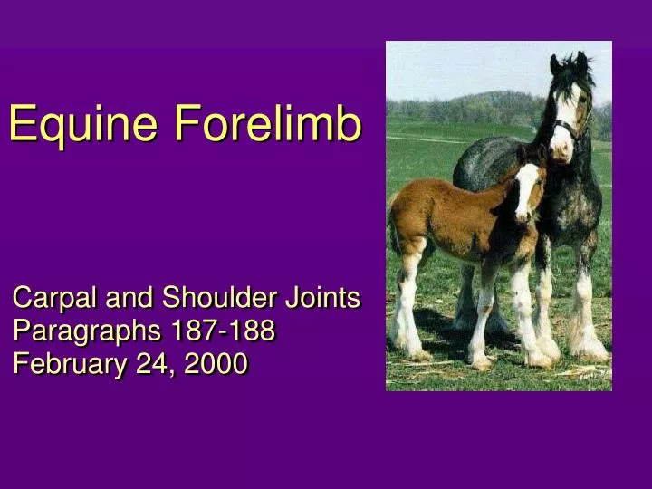
Equine Forelimb. Carpal and Shoulder Joints Paragraphs 187-188 February 24, 2000. Overview of February's Lecture Topics. Date PP Topic Feb. 9: 112-120 Shoulder and arm region; brachial plexus Feb. 10: 121-127 Vessels and nerves of the upper limb; intro to lower limb nerves

E N D
Equine Forelimb Carpal and Shoulder Joints Paragraphs 187-188 February 24, 2000
Overview of February's Lecture Topics Date PP Topic Feb. 9: 112-120 Shoulder and arm region; brachial plexus Feb. 10: 121-127 Vessels and nerves of the upper limb; intro to lower limb nerves Feb. 15: 128-136 Metacarpal structures; digital vessels and nerves; major arterial route of forelimb Feb. 16: 137-142 Forelimb bones and joints Feb. 17: 143-150 Stay apparatus and forelimb muscles; diagnostic nerve blocks; hoof and foot Feb. 22: 151-166 Hoof and foot; digital vessels and nerves; ligaments and joints of the manus Feb. 23: 167-186 Digit and suspensory apparatus Feb. 24: 187-188 Carpal and shoulder joints
Shoulder Joint • “Collateral ligaments” • there are no true collateral ligaments • muscle insertions perform this function • 1 major medial muscle - ? • 2 lateral muscles - ? and ? • Synovial structures • shoulder joint sac • bicipital bursa (no communication with above) • infraspinatus bursa
Bursae and Joint Sacs of the Shoulder and Elbow shoulder joint infraspinatus bursa (red) subtendinous olecranon bursa bicipital bursa subcutaneous olecranon bursa elbow joint (blue) lateral view
Carpal Ligaments lateral dorsal lateral collateral ligament medial collateral ligament ligaments of accessory carpal bone dorsal intercarpal ligaments dorsal carpometacarpal ligaments
Carpus - Review Bones http://cal.vet.upenn.edu/larad/index.html - radiology web site
Access to Carpal Joints (D553) lat. view
Dorsopalmar Radiographs X-Rays “free edges” - those that appear on the edge of the shadow. Can resolve those surfaces individually. Film Shadow
Break: Example Quiz Question • This joint is in • extension • flexion • hyperextension • T or F. Distension of the digital sheath may cause swelling in this area.
Lateromedial Radiographs X-Rays “free edges” - those that appear on the edge of the shadow. Can resolve those surfaces individually. Film
Oblique Radiographs • X-rays enter the carpus on the dorsolateral or dorsomedial aspect of the carpus. • The objective is to highlight particular bone surfaces that are not normally isolated with lateral or dorsopalmar views.
X-Rays Free edges seen on the film are the palmarolateral and dorsomedial edges. Film DLPM-O Carpal Radiograph DLPM-O: x-ray beams directed from dorsolateral toward palmaromedial. Note: accessory carpal and MCIV will be more easily visible in this view. Dorsomedial Dorsolateral Palmarolateral Palmaromedial
X-Rays Free edges seen on the film are the palmarolateral and dorsomedial edges. DLPM-O Carpal Radiograph Dorsomedial Palmarolateral
Example Quiz Question • Name the view. • Dorsolateral palmaromedial oblique • This is the (dorsomedial, dorsolateral) surface of the carpus. • Dorsomedial surface • This surface belongs to the (2nd, 3rd, 4th) carpal bone. • 3rd and 4th (superimposed) • Identify the bone. • Second metacarpal
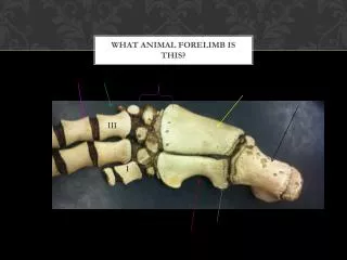
DLPM-O Carpal Radiograph dorsomedial aspect of carpus palmarolateral aspect of carpus accessory carpal (not superimposed) intermediate carpal ulnar carpal radial carpal fourth carpal second carpal third carpal MC IV (not superimposed) MC II MC III
X-Rays Film DMPL-O Carpal Radiograph DMPL-O: x-ray beams directed from dorsomedial toward palmarolateral. Dorsomedial Dorsolateral Note: accessory carpal is less easily observed in this view. Palmarolateral Palmaromedial Free edges seen on the film are the dorsolateral and palmaromedial edges.
X-Rays DMPL-O Carpal Radiograph Free edges seen on the film are the dorsolateral and palmaromedial edges. Dorsolateral Palmaromedial Film
DMPL-O Carpal Radiograph palmaromedial aspect of carpus dorsolateral aspect of carpus intermediate carpal accessory carpal ulnar carpal radial carpal third carpal second carpal fourth carpal MC II MC IV MC III