Download
1 / 59
610 likes | 914 Views
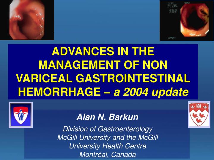
ADVANCES IN THE MANAGEMENT OF NON VARICEAL GASTROINTESTINAL HEMORRHAGE – a 2004 update. Alan N. Barkun. Division of Gastroenterology McGill University and the McGill University Health Centre Montr é al, Canada.

E N D
ADVANCES IN THE MANAGEMENT OF NON VARICEAL GASTROINTESTINAL HEMORRHAGE – a 2004 update Alan N. Barkun Division of GastroenterologyMcGill University and the McGill University Health Centre Montréal, Canada
Significant evolution in the management of patients with non variceal upper GI bleeding (supportive care, pharmacological treatment and endoscopic hemostasis) The last Consensus guidelines published: Gut 2002 – incomplete Before that: NIH Consensus Conference, almost 15 years ago INTRODUCTION
To review major advances in the management of patients with gastrointestinal hemorrhage To highlight the contribution of 2 major Canadian initiatives that have helped set the international standards of care: RUGBE The Banff Consensus Conference AIMS
Ann Int Med., 2003 – Banff Consensus group on Non Variceal Upper GI Bleeding
NON VARICEAL UPPER GASTROINTESTINAL BLEEDING ALL GI BLEEDERS Identify the high risk Pt 80% stop bleeding20% bleed on, ontheir own or re-bleed 20% RE-BLEEDING RATE TARGET GROUP ANY Rx
2484 procedures in 1869 patients • Endoscopy performed within 24 hrsin 76% other Other30% PUD56% Spurting 3% clean base46% Dieulafoy 2% oozing22% M-W tear 4% visiblevessel14% Esophagitis8% spot4% clot7% RUGBE: Endoscopic Findings Barkun et al., Am J Gastroenterol. 2004
Continued bleeding/rebleeding 14.1% Surgery 6.5% Mortality 5.4% Mean hospitalization 5.6±6.1 d Main Outcomes Barkun et al., Am J Gastroenterol. 2004
STATEMENT 1 Hospitals should develop institution specific protocols for multidisciplinary management, which should include access to an endoscopist with training in endoscopic hemostasis (III C) A: 100% 80% of RUGBE sites did not have a specific protocol
STATEMENT 2 Support staff trained to assist in endoscopy should be available for urgent endoscopy (III C) A: 92%, B: 8% Only 40% of all RUGBE sites had anurse taking availability call
STATEMENT 3 Immediate evaluation and appropriate resuscitation is critical to proper management (III C) A: 96%, B: 4% Recent level II data suggest that is true, but only historical control group (Baradarian Am J Gastro 2004)
STATEMENT 5 Clinical (non-endoscopic) stratification of patients into low- and high-risk categories for rebleeding and mortality is important for proper management. Available prognostic scales may be used to assist in decision making. (II-2 B) A: 76%, B: 24%
Blatchford criteria (BMJ, 1997) Adapted from BMJ 1997
Probability of Mortality Age = 65 Nb of comorbidities > 1 ASA > score 1 Bright blood per NGT = No Systolic blood pressure at initial assessment = 120 mm Hg yes 36.7 % yes Rebleeding 9.9 % no Bright blood per rectal exam 16.4 % yes no yes Rebleeding 3.6 % Inpatientsstatus at timeof bleeding no 17.3 % yes no Rebleeding yes 3.8 % Bright blood per rectal exam no 6.6 % yes no Rebleeding 1.3 % no Independent Predictors of MortalityClinical Scenarios(all patients who were not transferred)
STATEMENT 4 Inselected patients, the placement of a naso-gastric tube can be considered because the findings may have prognostic value (II-3 B) A: 42%, B: 33%, C: 25%
ROLE of NGA Aljebreen AM et al., 2003
STATEMENT 6 Early stratification of patients into low- and high-risk categories for rebleeding and mortality, based on clinical AND endoscopic criteria, is important for proper management. Available prognostic scales may be used to assist in decision making. (I A) A: 96%, B: 0, C: 4%
Rockall Score – Risk assessment of Death/Rebleeding (N=4185) RUGBE validation of the Rockall scoring has been submitted for publication (Enns et al.) Rockall, Lancet 1996
Allows for safe and prompt discharge of patients classified as low-risk* (I A) A: 92%, B: 8% Improves patient outcomes for patients classified as high-risk* (II-2 C) A: 64%, B: 36% Reduces resource utilization for patients classified as either low- or high-risk* (IA) A: 88%, B: 12% STATEMENT 7 Early endoscopy (within the first 24 hours) Increasing risk of negative outcome *by clinical and endoscopic criteria
Early endoscopy 75%
Prognostic Factors: Endoscopic Incidence of Re-bleeding by Appearance of Ulcer at Endoscopy Ia = spurter Ib = oozer Forrest IIb IIa Laine, Peterson, N Engl J Med 1994.
STATEMENT 8 Afinding of low-risk endoscopic stigmata (a clean based ulcer, or a non-protuberant pigmented dot in an ulcer bed) is not an indication for endoscopic hemostatic therapy (I A) A: 100%
STATEMENT 9 A finding of clot in an ulcer bed warrants targeted irrigation in an attempt at dislodgment. Endoscopic therapy for persistently adherent clots is controversial (III C -- Ia) A: 32%, B: 56%, C: 4%, D: 8% -- more unanimity
Active bleeding: “Spurter” (= trouble!)
STATEMENT 10 A finding of high-risk endoscopic stigmata (active bleeding or a visible vessel in an ulcer bed) is an indication for immediate endoscopic hemostatic therapy (I A) A: 100%
Meta-analysis (Cook, et al.Gastroenterol,1992) 30 trials (n=2,412) Similar results in an earlier meta-analysis of 25 trials (Sacks, et al. JAMA,1990) Treatments studied:thermal (laser), few injection, no combination or clips ORCI OR 95% CI Further Bleeding 0.38 0.32-0.45 Surgery 0.36 0.28-0.45 Mortality 0.55 0.40-0.76 OR=odds ratio for treatment vs. controls. Statistical heterogeneity was observed for bleeding and surgery. Endoscopic Therapy Recently confirmed by Bardou et al, 2003 (71 studies, 9000 patients)
STATEMENT 13 Monotherapy, with injection or thermal coagulation, is an effective endoscopic hemostatic technique for high-risk stigmata; however, the combination is superior to either treatment alone (I B) A: 36%, B: 48%, C: 16%
STATEMENT 14 The placement of clips is a promising endoscopic hemostatic therapy for high-risk stigmata (I B) A: 44%, B: 52%, C: 4%
STATEMENT 15 Routine second look endoscopy is not recommended (I E) A: 92%, B: 8%
STATEMENT 16 Incases of rebleeding, a second attempt at endoscopic therapy is generally recommended (I A) A: 100%
STATEMENT 19 Somatostatin and octreotide are not recommended in the routine management of patients (I C) ** A: 96%, B: 4%
100 Visible Vesselsn = 25-52 80 Adherent clot Visible vessel Active bleeding 60 Presence following endoscopic treatment on Day 0 40 25 13 9 9 20 8 8 6 5 0 0 Day 1 Day 2 Day 3 Early Risk of Re-bleedingNatural History of the Visible Vessel Most re-bleeding occurs withinthe first 72 hours % Lau et al, 1998
Acid is associated with Decreased platelet aggregation, and platelet disaggregation (in vivo, and animal models) – ideal pH approximately 6.5 Increased clot lysis due to pepsin activation by acid (in vitro) Increased fibrinolytic activity, that is impaired by acid suppression (in vitro, cell culture assays) Effect of acid suppression
STATEMENT 18 H2 receptor antagonists are not recommended in the management of patients (I D) A: 92%, B: 8%
Re-bleeding Surgery Mortality Absolute change (%) IV H2RA vs placebo * * o -3.2% -6.7% -7.2% NNT =14 NNT =15 NNT =32 Levine JA et al., APT, 2002 Effect of IV H2RA on Upper GI Bleeding:meta analysis of 1062 patients, 24 RCT’s NO differences in outcomes attributable to IV H2RA’s for ALL patients • Only significant differences amongst patients with bleeding gastric ulcers
Tolerance of H2RA Gastric pH Omeprazole IV Ranitidine IV Netzer, 1999
Role of PPI for UGI Bleeding:Continuous Infusion 3 RCT’s concur 1RCT “negative”* Lau, et al. NEJM 2000.
STATEMENT 20 An IV bolus followed by continuous infusion intravenous proton pump inhibitor is effective in decreasing rebleeding in patients who have undergone successful endoscopic therapy (I A) A: 100%
ci IV PPI vs placebo ci IV PPI vs H2RA Continuous Infusion IV PPI vs H2RA and placebo: meta-analysis Meta-analysis of 71 studies and over 9000 patients included 16 H2RA and 4 CI IVPPI studies since 1990 * 20% Absolute risk reduction in % in the model * 15.6% * 2.8% Bardou et al., submitted, 2004