Download
1 / 75
750 likes | 980 Views
Outline. Cell structureDNA to chromosomesCritical cellular targetCell cycleDNA strand breaksIntroduction of cell survival curves. Cells. Four concepts (collectively known as cell theory):the cell is the basic structural and functional unit of living organisms - defining cell properties defines lifethe activity of an organism is dependent on both the individual and collective activities of its cellsaccording to the principle of complementarity, the biochemical activities of cells are dete30088

E N D
1. Basic Cell Structure, Cycle and Division
- and -
DNA, Strand Breaks and Chromosomal Aberrations
(Travis & Prasad and Hall, Ch. 2)
2. Outline Cell structure
DNA to chromosomes
Critical cellular target
Cell cycle
DNA strand breaks
Introduction of cell survival curves
3. Cells Four concepts (collectively known as cell theory):
the cell is the basic structural and functional unit of living organisms - defining cell properties defines life
the activity of an organism is dependent on both the individual and collective activities of its cells
according to the principle of complementarity, the biochemical activities of cells are determined and made possible by specific subcellular structures
continuity of life has a cellular basis
The human body has ~ 50-60 trillion cells
4. Chemical Constituency of Cells Water
Proteins - amino acid chains
Carbohydrates - sugars, starches, etc.; Cx(H2O)
Nucleic acids - DNA, RNA
Lipids - fats
Salts - NaCl and KCl
6. Cytoplasm Cytoplasm
cellular material inside the plasma membrane and outside the nucleus
the site of most metabolic functions of the cell: anabolism (building up) and catabolism (breaking down) of organic compounds
consists of three major elements:
Cytosol - viscous semitransparent fluid that suspends the organelles within the cytoplasm
Organelles - membrane bound structures that compartmentalize the cytoplasm and allow the cell to operate in a highly organized manner
Inclusions - chemical substances such as lipid droplets or melanin grains (not present in all cells; not functioning units)
7. The Cell Nucleus Largest organelle (~ 5 mm dia.)
thicker membrane and more viscous fluid
Contains the nucleolus and chromatin
Gene-containing control center
the cell brain
regulates cellular processes
Most cells contain one nucleus, however, some are
anucleate (no nucleus) - red blood cells
cannot reproduce; die after a few months
multi-nucleate (several nuclei) - skeletal muscle cells
9. Plasma Membrane Composed of a lipid bi-layer with embedded proteins
membrane wall is hydrophobic (impermeable to water)
but, proteins embedded in cell wall allow for diffusion
Functions to control exchanges between the cell and the outside world
Selectively permeable structure that prohibits the passage of some substances and permits the passage of others
10. Lysosomes Spherical membranous bags containing digestive enzymes used to break down proteins, DNA and carbohydrates
enzymes are capable of digesting the cell if they are accidentally released
Function to:
digest particles ingested by endocytosis (bacteria, viruses)
remove nonfunctional organelles
break down stored hormones
Many agents capable of altering permeability of the lysosomal membrane (including radiation)
can result in the release of the enzymes
11. Golgi Complex Major function - to modify, concentrate, and package proteins/membranes
Vesicles, containing proteins destined for export, pinch off from the Golgi complex as secretory vesicles and migrate to the plasma membrane to discharge their contents from the cell by exocytosis
Not all functions are known
12. Mitochondria Energy producers for cellular functions
by breaking down nutrients through �oxidation�
also manufacturers of ATP (adenosine triphosphate; stores energy)
cell powerhouses
Carbohydrates - primary source of energy
Number in a cell depends on energy requirements
cardiac cells have many more than lymphocytes
Contain RNA and are self replicating organelles ATP - adenosine triphospate
DNA - deoxyribonucleic acid
RNA - ribonucleic acidATP - adenosine triphospate
DNA - deoxyribonucleic acid
RNA - ribonucleic acid
13. Endoplasmic Reticulum An extensive system of interconnected tubes and parallel membranes enclosing fluid-filled cavities
Tubules arranged in a branching network and containing enzymes to catalyze several reactions
Function varies with cell type
Two distinct varieties:
rough - (protein synthesis) external surface is studded with ribosomes
smooth - (not completely known) no ribosomes on surface
14. Ribosomes Cytoplasmic organelles made up of protein and ribosomal RNA
Synthesize cellular proteins
Free ribosomes
floating in the cytoplasm
produce soluble proteins that function in the cytosol
Membrane-bound ribosomes
those on the rough endoplasmic reticulum
synthesize protein products destined for cellular membranes or for export
15. RNA Ribonucleic acid
Found in nucleus and cytoplasm
Nuclear RNA - transmits genetic instructions from nucleus to the cytoplasm
Cytoplasmic RNA - functions in the assembly of proteins
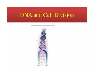
17. DNA DNA (deoxyribonucleic acid) is a coiled double-helical polymer (strands of proteins and sugars)
The base unit of DNA is called the nucleotide
composed of a deoxyribose-sugar molecules linked to a phosphate group (adenine, guanine, cytosine, thymine)
19. Levels of DNA condensation DNA double-strand helix.
Chromatin strand (DNA with histones).
Condensed chromatin during interphase with centromere.
Condensed chromatin during prophase. (Two copies of the DNA molecule are now present)
Chromosome during metaphase.
20. Chromosome details It is difficult to appreciate details of chromosome structure even with an electron microscope.
However, one label them with dyes that are preferentially taken up by certain regions
These modifications create a banding pattern that can be used to identify and characterize individual chromosomes.
21. DNA and Chromosomes To understand how the DNA and histones are organized in a chromosome, we must appreciate the fact that the nucleus is only 6 micrometers in diameter.
The total length of DNA in the human genome is 1.8 meters.
Thus, in order to pack the DNA into the nucleus as in the photograph of the metaphase chromosome , there must be several levels of coiling and supercoiling.
There is nearly a 10,000-fold reduction in length in an interphase nucleus.
Each chromosome contains 1 long molecule of DNA plus associated histones (basic proteins) which help in the condensation and regulation processes.
22. Chromosome Organization Different levels of uncoiling in the chromosome are shown.
4 nm DNA filaments are labeled the "DNA helix".
Double stranded DNA is wrapped around sets of 8 histones to form a 10 nm filament.
Sets of 8 histones wrapped by DNA
are separated by spacer regions of 4 nm DNA filament (double stranded DNA) and Histone H1.
are called "nucleosomes".
Nnext level of coiling produces 30 nm nucleoprotein fibers
Further looping of nucleoprotein fibers around a protein scaffold forms the individual metaphase chromosomes
23. Genes The unit of genetic material responsible for directing cytoplasmic activity and transmitting hereditary information
Each gene contains a finite section of DNA with specific base sequence coding
Each genes occupies a specific chromosomal locus
24. Chromosomes Each chromosome contains many genes arranged in a specific linear sequence
Constricted at certain points by a centromere, a clear region necessary for movement of the chromosome during cell division
Chromosomes are constant in number for each species
25. Relative Sizes of Genetic Materials
26. Critical Target There is strong evidence that DNA is the main target for biological effects of radiation
Some of this evidence comes from microbeam and microsurgery experiments where different parts of cells were irradiated and transplanted (more later)
27. DNA Components Adenine and guanine are purine-based (C5H4N4) components
Cytosine and thymine are pyrimidine-based (C4H4N2) components
28. DNA Structure Adenine pairs with thymine and guanine pairs with cytosine - always
29. Human Cells Two categories:
Somatic Cells
organs, tissues, structures, etc.
Germ Cells
those associated with reproduction
30. Somatic Cells Contain 2 sets (�diploid�) of 23 chromosomes
Mammalian cells proliferate by �mitosis�
In mitosis, one parent cell divides into two identical daughter cells
Both daughter cells receive a nearly equal portions of the cellular material
31. Chromosome Numbers
All animals have a characteristic number of chromosomes in their body cells called the diploid (or 2n) number.
These occur as homologous pairs, one member of each pair having been acquired from the gamete of one of the two parents of the individual whose cells are being examined.
The gametes contain the haploid number (n) of chromosomes
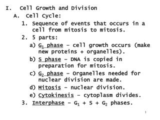
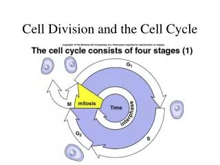
32. The Cell Life Cycle The series of changes a cell goes through from the time it is formed until it reproduces is its life cycle
The life cycle is comprised of two major periods:
Interphase
cell grows and carries on its usual activities
Mitosis (mitotic phase)
cellular reproduction
33. The Cell Cycle
34. Interphase The �growth phase�; preparing for next division
Total period from cell formation to cell division
During Interphase the chromosomal material is seen in the form of diffuse chromatin
Interphase is divided into G1, S, and G2 sub-phases:
G1 - (1st growth period) cells synthesize proteins and grow vigorously
S - (synthesis) replication of DNA
G2 - (2nd growth period) enzymes/proteins needed for division are synthesized and moved to their proper sites
35. DNA Replication S phase
Must occur before division, so that identical copies of the cell�s genes can be passed on
Process includes:
an enzyme uncoils, untwists and separates the DNA molecule into two complementary nucleotide chains
two identical strands of DNA result
each strand has half of the old DNA molecule and half is newly synthesized
36. Mitosis Cell division
Divided into four distinct phases:
prophase (early and late)
metaphase
anaphase
telophase (and cytokinesis)
37. Early Prophase Prophase is the first and longest phase of mitosis
Begins when the chromatin threads start to coil and condense, forming barlike chromosomes that are visible under a light microscope
Each chromosome consists of two identical chromatin threads, called chromatids, attached by a small button-like body called a centromere
The centriole pairs migrate to opposite poles of the cell
Mitotic spindles grow from the regions of the centrioles
38. Late Prophase Nuclear membrane then fragments
allowing the spindles to occupy the center of the cell and to interact with the chromosomes
Spindles attach to the centromeres at one end and are anchored to the polar regions of the cell at the other
chromosomes end up with spindles attached to them from both poles of the cell
Spindles tug on the centromeres and draw the chromosomes to the center of the cell
39. Metaphase Chromosomes line up in the center of the cell (forming the equatorial plate)
Nuclear membrane dissolves and chromosomes are free to move
The two chromatids of each chromosome are attached to the mitotic spindle at their centromere
40. Anaphase Centromere splits and each chromatid becomes a chromosome
Chromosomes are gradually pulled toward the opposite poles of the cell
Cell elongates considerably
Duplicate chromosomes are now located at the opposite poles of the cell
Typically lasts only a few minutes (shortest phase)
41. Transition from Metaphase to Anaphase; Chromosomes Split
42. Telophase Essentially prophase in reverse
Chromosomes at opposite ends of the cell uncoil and resume their threadlike extended chromatin form
A new nuclear membrane, derived from the rough endoplasmic reticulum, reforms around each chromatin mass
For a brief moment the cell is binucleate, with two identical nuclei
43. The role of Telomeres Cap and protect end of DNA
Long arrays of TTAGGG,
1.5 � 150 kbases
At each division telomeric DNA lost
After ~40-60 divisions, cap is lost and cell dies (senesces)
Called a �molecular clock�
Stem & cancer cells avoid problem by rebuilding chromosome ends
Activate enzyme telomerase.
44. Cytokinesis As mitosis draws to a close, cytokinesis (physical cell division) occurs, and the cell divides into two daughter cells
The cytoplasm and organelles are evenly distributed between the two new daughter cells
45. Germ Cells Germ cells are produced by organisms for the sole purpose of sexual reproduction
Oogenesis - the process of germ-cell production in the female
leads to the development of an ovum
Spermatogenesis - the process of germ-cell production in the male
leads to the production of spermatozoa
46. Meiosis The process by which germ cells divide
Germ cells contain only 1 (�haploid�) set of 23 chromosomes
Meiotic division similar to mitotic division
Exceptions:
no DNA replication
daughter cells have only half of the genetic material of the parent cell
47. The Common Theory of Biological Damage Resulting from Radiation Exposure
48. Review - Energy Loss by charged particles Heavy charged particles lose kinetic energy via a sequence of small energy transfers to atomic electrons in the medium.
Most energy deposition occurs in the infratrack, a narrow region around the particle track extending about 10 atomic distances.
Ionization outside the infratrack is caused by very energetic particles that escape from the infratrack and secondary electrons.
The more energetic interactions eject electrons from their parent atoms and generate primary ion-pairs.
An approximate expression for the maximum energy transfer to an electron from a heavy charged particle of mass number A and energy E (MeV) is given by:
Wmax = 215 E /A
where Wmax is in eV.
Thus secondary electrons generated by a 5 MeV alpha particle range up to about 300 eV of kinetic energy.
49. Energy Loss by charged particles Energetic secondary e�s can initiate additional ionizations, while less energetic ones induce electronic excitations.
Lowest energy secondary e�s are referred to as "sub-excitation", whose role in biological radiation damage remains unclear.
Only a small fraction of initial energy is transferred at each event, a track consisting of clusters of ions or spurs is generated along the path of the moving particle.
Most spurs in water comprise 1-5 ion-pairs.
These tracks may be visualized in a cloud chamber by their vapor trail.
High-energy secondary electrons are occasionally generated. Energy loss by these energetic electrons leads to short branching tracks or "delta rays"
Delta rays may terminate in larger pear-shaped regions of ionisation or "blobs".
Similar considerations apply for energy transfer to a fluid medium in indirect action.
50. Energy Loss by charged particles Spurs and excitations in the track of an alpha-particle in water.
Each circle depicts an ionization or excitation event.
The branching tracks are "delta rays".
51. Distribution of Ion-pairs in water from passage of fast electrons and beta particles Fast electrons & betas lose energy by inelastic collisions with electrons of the medium.
Electrons tracks are less dense than the tracks of heavy charged particles, owing to the lower LET, and the spurs are more widely spaced with more frequent delta rays terminating with blobs.
Only about 20% of beta particles penetrate to the maximum range owing to their broad energy distribution.
In addition to energy deposition, electrons undergo elastic electron-electron collisions leading to multiple scattering and curvature of the tracks which complicate the dosimetry in extended sources
52. Ion pair creation in water from 20 Electrons
53. DNA Strand Breaks DNA can suffer single and double strand breaks
Strong circumstantial evidence that DNA is principal target for cell killing
A single-strand break occurs when only one of the helices suffers a break
A double-strand break occurs when both helices suffer a break
either directly opposite one another or when separated by only a few base pairs
54. Diagram of single and double strand DNA breaks
55. Diagram of single and double strand DNA breaks
56. Diagram of single and double strand DNA breaks
57. Diagram of single and double strand DNA breaks
58. Single-Strand Breaks Readily repaired
usually does not lead to mutations or cell death
Improper repair is possible
normally leads to mutations or death
59. Double-Strand Breaks Difficult or impossible to repair
may lead to programmed cell death (apoptosis), mutation or carcinogenesis
If not repaired, or repair is in error, mutations may be replicated
may lead to cells that function improperly and have unregulated cell growth (e.g., cancer)
60. Measuring DNA Strand Breaks Single- and double-strand breaks of DNA can be readily measured by using ordinary DNA �finger-printing� techniques
The DNA is isolated and processed to analyze the location and nature of the breaks
61. Fragment Behavior (post-break) Repair
breaks may rejoin in their original configuration
Aberration
breaks may fail to rejoin
broken ends may rejoin other broken ends, etc
occurs at next mitosis
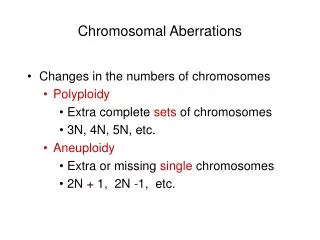
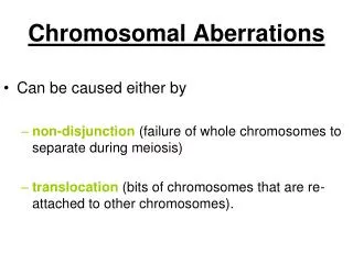
62. Chromosomal Aberrations Often caused by breakage and incorrect rejoining
broken segments may remain separated from minutes to hours; ends are said to be �sticky�
they are capable of reattaching to any other broken segments (most often rejoining in their original configuration)
breaks during specific phases of mitosis result in different endpoints (as we will see)
63. Radiation-Induced Aberrations Occur when cell is irradiated before the chromosome material has been duplicated
Frequency of single-strand breaks increases linearly with radiation dose
Frequency of double-strand breaks increases with dose as a power function (power of ~1.5 to 2)
64. Radiation-Induced Aberrations Dose-rate effect for single-strand breaks
lower dose rates allow for greater probability of repair
provided there is sufficient time for the single-strand break to be repaired prior to the formation of a double-strand break in the vicinity
thus, two neighboring single-strand breaks (identical to a double-strand break) less likely
possibility of a �threshold� dose rate
65. Radiation-Induced Aberrations 3 major lethal aberrations
dicentric
ring
anaphase bridge
2 major non-lethal aberrations
translocation
deletion
66. Example: Dicentric (lethal) Steps in the formation of a dicentric and an acentric fragment
67. Example: Ring (Lethal) Steps in the formation of a chromosomal ring
68. Example: Bridge (Lethal) Steps in the formation of an anaphase bridge and an acentric fragment
69. Rearrangements Not lethal; involved in carcinogenesis
translocation
breaks in two chromosomes
the sticky ends are exchanged
deletion
two breaks in one chromosome
information between the two breaks is lost
70. Symmetric Translocation (non lethal) Pre-replication chromosomes
Radiation induces breaks in adjacent chromosomes
Broken pieces exchanged
Not necessarily lethal to cell
May lead to cancer because of loss of suppressor gene (in fragment)
71. Small Interstitial Deletion (non lethal) Pre-replication chromosomes
Radiation induces adjacent breaks in chromosome
Fragment lost at next mitosis
Not necessarily lethal to cell
May lead to cancer because of loss of suppressor gene (in fragment)
72. Implications Potential for some aberrations to lead to disease, i.e. cancer
Specific translocations have been associated with several human malignancies
Non-lethal aberrations can be detected in irradiated persons for up to 40 years after exposure
biological dosimeters
73. Implications The formation of a dicentric, ring, or bridge usually leads to cell death
�Cell survival curves� are used to quantify the effect
74. Frequency of Chromosomal Aberrations Linear-quadratic function of dose
Aberrations result from 2 separate breaks
75. Cell Survival Curve The curve is characterized by two regions:
linear region (aD)
double-strand break from a single entity
the probability of this single event is proportional to dose (D)
quadratic region (bD2)
two, single-strand breaks from two different entities
the probability of these two events is proportional to dose * dose (D2)
76. more about cell survival curves next time �.