Download
1 / 37
370 likes | 476 Views
Transport/Circulatory System. Blood Heart Circulation Lymphatics Immunity. Human Circulatory System. Blood Heart Arteries Arterioles Capillaries Venules Veins Lymph Vessels Lymph Fluid. Functions of Blood. 3 Major Functions of Blood are… Transport Regulation Protection.

E N D
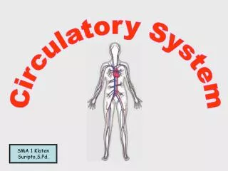
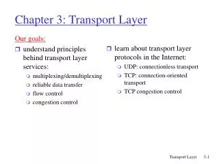
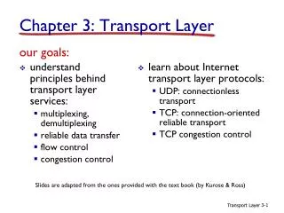
Transport/Circulatory System Blood Heart Circulation Lymphatics Immunity
Human Circulatory System Blood Heart Arteries Arterioles Capillaries Venules Veins Lymph Vessels Lymph Fluid
Functions of Blood 3 Major Functions of Blood are… • Transport • Regulation • Protection
Blood Composition Blood consists of 2 main parts… • Plasma (55%) • Cells (Red Blood Cells, White Blood Cells, & Platelets) (45%)
Blood Composition • Plasma (55%) • Amber coloured fluid (92%) • Dissolved materials (8%) • Nutrients (sugars, amino acids, vitamins) • Gases (O2 & CO2) • Hormones (Chemical Messengers) • Antibodies (infection fighters) • Proteins (eg. Prothrombin) • Salts (Sodium Chloride, Bicarbonate) • Wastes (eg. Urea, Heat)
Blood Composition 2. Cells (45%) • Red Blood Cells • Red blood cells (RBC) make up most of the cellular part of blood. • White Blood Cells • defenders of the blood circulatory system. • Platelets • involved in clotting (initiate clotting process)
RBCs Red Blood Cells aka. Erythrocytes (“erythro” meaning red in Greek) Physical Appearance • Small (~ 8 m in diameter) • Biconcave Disks
RBCs • No Nucleus • Very Numerous • 3 – 4 month lifespan • Dead RBCs broken down by liver • New RBCs are produced by bone marrow • Contains hemoglobin (iron rich pigment)
RBCs Function • Carry O2 and CO2 • O2 + Hemoglobin = Oxyhemoglobin • CO2 + Hemoglobin = Carbaminohemoglobin • Hemoglobin will also carry Carbon monoxide
RBCs Disorder : Anemia • Not enough RBCs in the blood Symptoms • Fatigue • Listlessness • Increased susceptibility to other diseases Treatment • Rest • Increased Iron intake
RBCs Sickle Cell Anemia Caused by a Mutant Gene producing defective hemoglobin Results in RBCs curving like a sickle Symptoms of anemia due to defective hemoglobin and not enough oxygen being carried to the cells
Sickle Cell Anemia Abnormal cells tend to form clumps and clog smaller blood vessels causing decrease in circulation… • Severe pain in abdomen, back, head, and extremities • Enlargement of heart, atrophy in brain cells • Cells die (hemolyze) easily resulting in severe anemia • Victims tend to suffer early death Evolutionary Benefits • People who have heterozygous state suffer only slight symptoms, but have a resistance to malaria
WBCs White Blood Cells aka. Leukocytes Function • Defend the body against foreign invaders
WBCs • Have a Nucleus • ~1 WBC to every 600 RBCs Physical Appearance • larger than RBCs (~10 µm) • generally round, but can change shape
WBCs Disorder : Leukemia • Cancer of blood forming organs • Increase in WBCs • Decrease in RBCs, results in Anemia • Immense number of WBCs do not mature Treatment • Cancer Treatments • Bone marrow transplant
WBCs Granulocytes • Formed in bone marrow • granules in the cytoplasm • Irregular-shaped nuclei • Short-lived
WBCs Neutrophils • ~65% of WBCs • actively phagocytic • engulfs foreign invaders
WBCs Eosinophils • ~ 2-4% of WBCs • Destroy foreign proteins • Break up blood clots
WBCs Basophils • ~ 0.5% of WBCs • Contains Histamine • Initiates swelling • Contains Heparin • Anticoagulant
WBCs Lymphocytes • Makes up 20-25% of WBCs Two types • B-Cells • Forms antibodies • T-Cells • Memory Storage
WBCs Monocytes • ~3-8% of WBCs • Actively phagocytic • Macrophages - can eat up to 100 bacteria at a time
Platelets Platelets • Smaller than RBCs • ~3 µm in diameter Contains • Thromboplastin • Serotonin
Platelets Disorder : Hemophilia • Affects mostly males • Inability to form blood clots Cause • Genetic • Mutant gene codes for defective protein Treatment • Injections of missing protein
Blood – “Clotting Cascade” If a blood vessel is damaged… • Platelets are fragile cells, when they hit a part of damaged wall (torn vessel), they break open. • Serotonin (hormone) is released, causing vasoconstriction • Thromboplastin (protein) is released, activating prothrombin (plasma protein). • Prothrombin Thrombin
Blood – “Clotting Cascade” • Thrombin reacts with fibrinogen causing the formation of fibrin (fibers) • Fibrin mesh traps RBCs • Mesh + RBCs = Blood Clot (Thrombus)
Dangers of Blood Clotting • Blood clots prevent the passage of blood • Area tissues do not get oxygen • If occurs in brain stroke • If occurs in heart vessel may have heart attack • A dislodged clot in vessel: embolus • May get caught in a vessel in a vital organ, causes embolism (coronary, pulmonary)
Blood - What’s Your Type? Historically, sometimes blood transfusions would keep people alive, other times it wouldn’t. Why? Karl Landsteiner found that different blood types existed. Glycoproteins on RBCs determine blood type: • Type A, B, AB, or O • Glycoproteins (A or B) are called antigens • Type O has no glycoproteins • Type AB has both A and B
Blood – Antigen-Antibody Response • How does the body recognize blood type? • Body has antibodies for the other antigen (e.g. Type A person will have Anti-B antibodies) • Antibodies respond to invaders by binding to surface proteins • With blood, RBC of a different type will agglutinate, or clump.
Blood - To whom can I donate? • Successful transfusion can occur provided the plasma of the patient and the erythrocytes of the donor are compatible. For example… Kevin (Type A) can donate to Michelle (Type A) and Donnie (Type AB)
Blood - To whom can I donate? A person with what blood type would be considered a Universal Donor? Type O Why? A person with what blood type would be considered a Universal Recipient? Type AB
Rhesus Factor – The Monkey Factor • A different blood antigen • Genetically determined • Two types: Rh- and Rh+ ~ 83.3% of population are Rh+ (protein present) ~ 16.6% of population are Rh– (protein absent) • Rh- will develop the Rh antibody only when exposed to Rh+ blood • Blood donating: Rh+ can receive from Rh-, but Rh- cannot receive from Rh+
Rhesus Factor – The Monkey Factor Important for pregnant women: • If there is a tear in the placenta, babies and mom’s blood cells can enter each other’s blood stream • If mother is Rh- and baby is Rh+, the babies RBC will stimulate the production of Rh antibodies by the mother • Erythroblastosis fetalis – If the mother’s Rh antibodies enter the babies blood stream, they will cause agglutination and destruction of babies RBCs, resulting in death of the baby. Treatment • Immunization of the mother with “Rhogam” or “Wingam”- medicines that prevent the formation of Rh antibodies (contain Rh anti-antibodies)