Download
1 / 1
10 likes | 107 Views
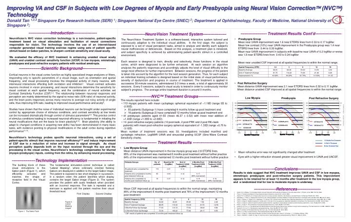
First Display. Second Display. Low Myopia. Presbyopia. Post Refractive Surgery. UCVA=20/54. UCVA=6/15. UCNVA=6/13. UCNVA=20/54. UCVA=6/13. UCVA=20/44. UCVA=6/7.5. UCVA=20/28. UCNVA=6/8. UCNVA=20/35. UCVA=20/25. UCVA=6/7.5. UCVA=6/8. UCVA=20/30. Spatial Frequency.

E N D
First Display Second Display Low Myopia Presbyopia Post Refractive Surgery UCVA=20/54 UCVA=6/15 UCNVA=6/13 UCNVA=20/54 UCVA=6/13 UCVA=20/44 UCVA=6/7.5 UCVA=20/28 UCNVA=6/8 UCNVA=20/35 UCVA=20/25 UCVA=6/7.5 UCVA=6/8 UCVA=20/30 Spatial Frequency Spatial Frequency Spatial Frequency Spatial Frequency Spatial Frequency Spatial Frequency Spatial Frequency Spatial Frequency Spatial Frequency Spatial Frequency Spatial Frequency Spatial Frequency Improving VA and CSF in Subjects with Low Degrees of Myopia and Early Presbyopia using Neural Vision Correction (NVC) Technology Donald Tan 1,2,3 Singapore Eye Research Institute (SERI) 1; Singapore National Eye Centre (SNEC) 2; Department of Ophthalmology, Faculty of Medicine, National University of Singapore 3; • -----------------Introduction------------------ • NeuroVision’s NVC vision correction technology is a non-invasive, patient-specific treatment based on visual stimulation and facilitation of neural connections responsible for vision. The technology involves the use of an internet-based computer generated visual training exercise regime using sets of patient specific stimuli based on Gabor patches, to sharpen contrast sensitivity and visual acuity. • We evaluated the efficacy of NVC treatment in enhancing unaided visual acuity (UAVA) and unaided contrast sensitivity function (UCSF) in low myopes, emmetropic presbyopes and post-refractive surgery patients with residual ametropia. • -------------------- Treatment Results Cont’d-------------------- • Presbyopia Group Mean near UAVA improvement was 1.6 near ETDRS lines from 0.33 to 0.17 logMar Mean low contrast (10%) near UAVA improvement in the Presbyopia group was 1.4 near ETDRS lines from 0.44 to 0.30 logMar. Mean near UAVA improvement in subjects with baseline near UAVA of 0.2 logMar or worse was 1.9 near ETDRS lines from 0.43 to 0.24 logMar • Mean near unaided CSF improved at all spatial frequencies to within the normal range. • Post Refractive Surgery • Mean distance UAVA improvement was 2.1 near ETDRS lines from 0.32 to 0.11 logMar Mean distance unaided CSF improved at all spatial frequencies to within the normal range. • Mean refractive error was not significantly changed after treatment • Eyes with a higher refraction showed greater visual improvement in UAVA and UACSF. -----------------NeuroVision Treatment System------------------ The NeuroVision Treatment System is a software-based, interactive system tailored and continuously adaptive to the individual visual abilities. In the first stage, the subject is exposed to a set of visual perception tasks, aimed to analyze and identify each subject’s neural inefficiencies or deficiencies. Based on this analysis, a treatment plan is initialized, and subject specificity is achieved by administering patient-specific stimuli in a controlled environment. Each session is designed to train, directly and selectively, those functions in the visual cortex, which were diagnosed to be further enhanced. At each session an algorithm analyzes the patient's responses and accordingly adjusts the level of visual difficulty to the range most effective for further improvement. Between sessions, the progress of the patient is taken into account by the algorithm for the next session generation. Thus, for each subject an individual training schedule is designed based on the initial state of visual performance, severity of dysfunction and progress in course of treatment. The treatment is applied in successive 30-minute sessions, administered 2-3 times a week, a total of approximately 30 sessions. Every 5 sessions, subject’s visual acuity is tested in order to continuously monitor subject’s progress. The average entire treatment duration is around 3 months. -----------------Scientific Background------------------ Cortical neurons in the visual cortex function as highly specialized image analyzers or filters, responding only to specific parameters of a visual image, such as orientation and spatial frequency, and visual processing involves the integrated activity of many neurons, with interneural interactions effecting both excitation and inhibition1. Visual contrast activates neurons involved in vision processing, and neural interactions determine the sensitivity for visual contrast at each spatial frequency, and the combination of neural activities set Contrast Sensitivity Function (CSF)1,2. The relationship between neuronal responses and perception are mainly determined by the signal-to-noise ratio (S/N ratio) of neuronal activity, and the brain pools responses across many neurons to average out noisy activity of single cells, thus improving S/N ratio, leading to improved visual performance and acuity3. Studies have shown that the noise of individual neurons can be brought under experimental control by appropriate choice of stimulus conditions, and contrast sensitivity at low levels can be increased dramatically through control of stimulus parameters4-8. This precise control of stimulus conditions leading to increased neuronal efficiency is fundamental in initiating the neural modifications that are the basis for brain plasticity9,10. Brain plasticity (the ability to adapt to changed conditions in acquiring new skills) has been demonstrated in many basic tasks, with evidence pointing to physical modifications in the adult cortex during repetitive performance11-12. NeuroVision’s technology probes specific neuronal interactions, using a set of patient-specific stimuli that improve neuronal efficiency6,13 and induce improvement of CSF due to a reduction of noise and increase in signal strength. As visual perception quality depends both on the input received through the eye and the processing in the visual cortex, NeuroVision’s technology compensates for blurred (myopic/presbyopic) inputs, coming from the retina, by enhancing neural processing. • -----------------NVC Treatment Groups---------------- • The results reported here include: • 113 myopic patients with mean cycloplegic spherical equivalent of –1.18D (range 0D to -2.50D) • 55 patients (Subgroup-1) have completed 6 months follow up post treatment end • 16 patients (Subgroup-2) have completed 12 months follow up post treatment end • 41 presbyopic patients aged 41-55 (mean 46.37 ± 0.52) with mean near addition of +1.40D (range +1.00D to +2.00D) • 14 post-refractive surgery patients (10 post-lasik, 2 post-PRK and 2 post RK+lasik patients) with mean post-refractive surgery spherical equivalent of -1.33D (range +0.75 to -3.12D) • Mean number of treatment sessions was 33. Investigations included manifest and cycloplegic refraction, LogMAR UAVA and sinusoidal grating UCSF (SineWave Contrast Sensitivity charts). • ----------------------- Treatment Results -------------------------- • Low Myopia Group Mean distance UAVA improvement in the low myopia group was 2.8 ETDRS lines. 85% of the improvement was maintained 6 months post treatment without further practice 84% of the improvement was maintained 12 months post treatment without further practice • Mean CSF improved at all spatial frequencies to within the normal range, maintaining 85% of the improvement 6 months post treatment and 76% of the improvement 12 months post treatment • * CSF results of patients who completed 12 months post treatment follow up ------------------Technology Implementation ------------------- The building block of these visual stimulations is the Gabor patch (Figure 1), which efficiently activates and matches the shape of receptive field in the Visual Cortex. The fundamental stimulation-control technique is called “Lateral Masking”, where collinearly oriented flanking Gabors are displayed in addition to the target Gabor image. The patient is exposed to two short displays in succession, in a random order; the patient identifies which display contains the target Gabor image (Figure 2). The system provides the patient with audio feedback when provided with an incorrect response. The task is repeated and a staircase is applied until the patient reaches their visual threshold level. -----------------Conclusions------------------ Results to date suggest that NVC treatment improves UAVA and CSF in low myopes, emmetropic presbyopes and post-refractive surgery patients. This improvement appears to be retained for at least 12 months after treatment in the low myopia group, and a randomized trial for low to moderate myopia is underway. -----------------References------------------ Hubel, D. H. & Wiesel, T. N. Receptive fields, binocular interaction and functional architecture in the cat's visual cortex. J. Physiol. (Lond.)160, 106-154 (1962). Polat, U. Functional architecture of long-range perceptual interactions. Spat Vis12, 143-62 (1999). Geisler, W. S. & Albrecht, D. G. Visual cortex neurons in monkeys and cats: detection, discrimination, and identification. Vis Neurosci14, 897-919 (1997). Kasamatsu, T., Polat, U., Pettet, M. W. & Norcia, A. M. Colinear facilitation promotes reliability of single-cell responses in cat striate cortex. Exp Brain Res138, 163-72. (2001). Polat, U., Mizobe, K., Pettet, M. W., Kasamatsu, T. & Norcia, A. M. Collinear stimuli regulate visual responses depending on cell's contrast threshold. Nature391, 580-4 (1998). Polat, U. & Sagi, D. Spatial interactions in human vision: from near to far via experience- dependent cascades of connections. Proc Natl Acad Sci U S A91, 1206-9 (1994). Polat, U. & Sagi, D. Lateral interactions between spatial channels: suppression and facilitation revealed by lateral masking experiments. Vision Res33, 993-9 (1993). Polat, U. & Sagi, D. The architecture of perceptual spatial interactions. Vision Res34, 73-8 (1994). Dosher, B. A. & Lu, Z. L. Perceptual learning reflects external noise filtering and internal noise reduction through channel reweighting. Proc Natl Acad Sci U S A95, 13988-93. (1998). Dosher, B. A. & Lu, Z. L. Mechanisms of perceptual learning. Vision Res39, 3197-221. (1999). Sagi, D. & Tanne, D. Perceptual learning: learning to see. Curr Opin Neurobiol4, 195-9 (1994). Gilbert, C. D. Adult Cortical Dynamics. Physiological Reviews78, 467-485 (1998). Polat, U. & Sagi, D. in Maturational Windows and Adult Cortical Plasticity (eds. Julesz, B. & Kovâcs, I.) 1-15 (Addison-Wesley, 1995). Polat, U., Ma-Naim, T. Belkin, M. Sagi, D. Improving vision in adult amblyopia by perceptual learning. PNAS 101, 6692-97 (2004). Figure 2: Lateral Masking images Figure 1: The Gabor Patch