Download
1 / 54
540 likes | 773 Views
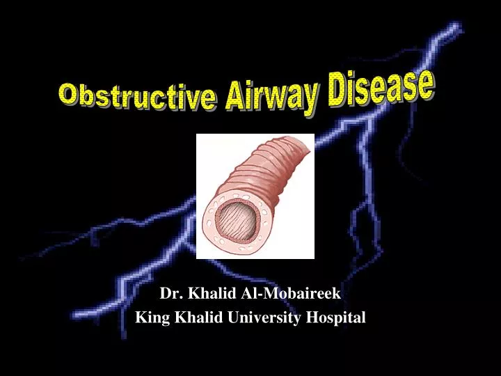
Obstructive Airway Disease. Dr. Khalid Al-Mobaireek King Khalid University Hospital. Obstructive airway Disease:. Reversible = Asthma Irreversible: Bronchiectasis Localized: Anatomical Airway: Internal, External, Parynchymal Diffuse: Aspiration Mucociliary clearance: PCD, CF

E N D
Obstructive Airway Disease Dr. Khalid Al-Mobaireek King Khalid University Hospital
Obstructive airway Disease: • Reversible = Asthma • Irreversible: Bronchiectasis • Localized: • Anatomical • Airway: Internal, External, • Parynchymal • Diffuse: • Aspiration • Mucociliary clearance: PCD, CF • Immune deficiency • Congenital • Post-infectious: Pertusis, TB, adenovirus..
Definition of Asthma • A chronic inflammatorydisorder of the airways • Many cells and cellular elements play a role • Chronic inflammation is associated with airway hyperresponsiveness that leads to recurrent episodes of wheezing, breathlessness, chest tightness, and coughing • Widespread, variable, and often reversible airflow limitation
Bronchospasm Edema, Mucus Hyperresponsiveness INFLAMMATION
Source: Peter J. Barnes, MD Asthma Inflammation: Cells and Mediators
Asthma Inflammation: Cells and Mediators Source: Peter J. Barnes, MD
NORMAL ASTHMA
AIR TRAPPING INSP EXP
Burden of Asthma • Asthma is one of the most common chronic diseases worldwide with an estimated 300 million affected individuals • Prevalence increasing in many countries, especially in children • A major cause of school/work absence
Asthma Prevalence 10 - 15%
Qaseem 13% Khobar 6% Riyadh 10 % Jeddah 13% Abha 17%
Factors that Influence Asthma Development and Expression • Host Factors • Genetic - Atopy - Airway hyperresponsiveness • Gender • Obesity • Environmental Factors • Indoor allergens • Outdoor allergens • Occupational sensitizers • Tobacco smoke • Air Pollution • Respiratory Infections • Diet
Environmental Allergens and Childhood Asthma • Dust mites • Furry pets • Molds • Cockroaches • Cigarette Smoking
History • Symptoms (cough, wheeze, SOB) • Onset, duration, frequency and severity • Activity and nocturnal exacerbation • Previous therapy • Triggers • Other atopies • Family history • Environmental history, SMOKING • Systemic review
Physical Examination • Growth parameter • ENT • Features of atopy • Chest findings • PEF
Investigations • Pulmonary Function Test • Chest X ray in some. • Allergy testing in some
Differential Diagnosis • Infections • Congenital Heart Disease • Foreign body • GER • Bronchopulmonary dysplasia • Structural anomalies
LEVEL OF CONTROL TREATMENT OF ACTION REDUCE maintain and find lowest controlling step controlled consider stepping up to gain control partly controlled uncontrolled step up until controlled INCREASE exacerbation treat as exacerbation REDUCE INCREASE TREATMENT STEPS STEP 1 STEP 2 STEP 3 STEP 4 STEP 5
Treatment objectives • Achieve and maintain control of symptoms • Maintain normal activity levels, including exercise • Maintain pulmonary function as close to normal levels as possible • Prevent asthma exacerbations • Avoid adverse effects from asthma medications • Prevent asthma mortality GINA Guidelines 2006
Treatment strategy 1. Develop Patient/Doctor Partnership 2. Identify and Reduce Exposure to Risk Factors 3. Assess, Treat and Monitor Asthma 4. Manage Asthma Exacerbations 5. Special Consideration GINA Guidelines 2006
Pharmacological therapy • Relievers • Inhaled fast-acting 2-agonists • Inhaled anticholinergics • Controllers • Inhaled corticosteroids • Inhaled long-acting 2-agonists • Inhaled cromones • Oral anti-leukotrienes • Oral theophyllines • Oral corticosteroids
Why don’t patients comply with treatment? • Intentional • Feel better • Fear of side effects • Don’t notice any benefit • Fear of addiction • Fear of being seen as an invalid • Too complex regimen • Can’t afford medication • Unintentional • Forget treatment • Misunderstand regimen / lack information • Unable to use their inhaler • Run out of medication
Cromolyn Sodium • Non-steroidal anti- inflammatory • Weak action on Early and late phases • Slow onset of action • If no response in 6 weeks change to ICS • Side effects: Irritation
Inhaled Corticosteroids • Effective in most cases • Safe especially at low doses • The anti-inflammatory of choice in asthma
Inhaled Steroids Side Effects • Growth: No significant effect at low to moderate doses. • Bones: not important • HPA axis: No serious clinical effect (high doses) • Alteration of glucose and lipid metabolism: Clinical significant is unclear (high doses) • Cataract: No increase risk • Skin: Purpura, easily bruising, dermal thinning • Local side effects
MANAGEMENT OF ACUTE ASTHMA
Assessment: History • Symptoms • Previous attacks • Prior therapy • Triggers
Physical examination: Signs of airway obstruction: • Fragmented speech • Unable to tolerate recumbent position • Expiration > 4 seconds • Tachycardia, tachypnea and hypotension • Use of accessory muscles • Pulsus paradoxus > 10 mmhg • Silent hyperinflated chest • Air leak
Physical examination: Signs of tissue hypoxia: • Cyanosis • Cardiac arrhythmia and hypotension • Restlessness, confusion, drowsiness and obtundation
Physical examination: Signs of Respiratory muscles fatigue: • Increase respiratory rate • Respiratory alterans (alteration between thoracic and abdominal muscles during inspiration) • Abdominal paradox (inward movement of the abdomen during inspiration)
Investigations: • Peak expiratory flow rate • Pulse oxymetry • ABG • CXR X ONLY IN FEW CASES
Oxygen • Hypoxemia is common • It worsens airway hyperreactivity • Monitor saturation
Inhaled β2 agonist Every 20 minutes in the first hour Assess after each nebulizer