Download

1 / 36
360 likes | 433 Views
Cellular Architecture . Or the typical/nontypical cell. Figure 4-1 Page 68. I. Limits to cell size . Surface to volume ratio 1. function of membrane 2. relationship of surface area to volume 3. consequences of growing too large. 1 mm. 2 mm. 2 mm. 1 mm. Figure 4-2 Page 68.

E N D
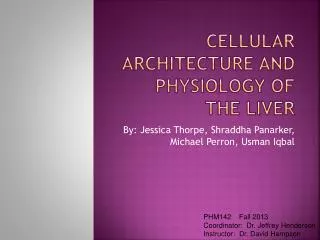
Cellular Architecture Or the typical/nontypical cell
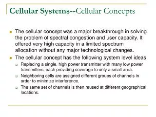
I. Limits to cell size • Surface to volume ratio 1. function of membrane 2. relationship of surface area to volume 3. consequences of growing too large
1 mm 2 mm 2 mm 1 mm Figure 4-2Page 68
I. Limits to cell size • Control issues • Informational flow • Transcription • Diffusion • Translation • Cell gets too large, lag time in the control of activities • Analogy
II. Few Generalizations • A. Procaryotic vs. eukaryotic • B. Typical cell • 1. animal • 2. plant • 3. discuss the similarities first
III. Organelles of synthesis • A. Introduction • 1. Going to act as an assembly line • 2. Nucleus • 3. Endoplasmic reticulm • 4. ribosome • 5. Golgi apparatus
III. Organelles of synthesis • Nucleus-headquarters 1. chromatin 2. chromosomes 3. nucleolus 4. double membrane with pores
III. Organelles of synthesis • C. Endoplasmic Reticulum • 1. definition • 2. nickname • 3. types of • Rough • Smooth
III. Organelles of synthesis • D. Ribosomes-protein synthesis • 1. found free in cytoplasm-endogenous use • 2. attached to E.R.-proteins for export • 3. responsible for translation of mRNA into protein
III. Organelles of synthesis • D. Golgi Body or Golgi Apparatus • 1. nick name of bottling center • 2. modifies product • 3. concentrates product • 4. packages product into vesicles • 5. exports via exocytosis
III. Organelles of synthesis • E. Cell membrane with exocytosis
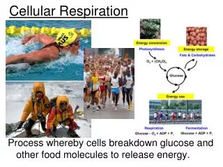
IV. Organelles of homeostasis • A. Mitochondria • Nickname • Structure • Endosymbiosis • More active tissue • Both animal and plant
B. Chloroplasts • Nickname • Structure • Endosymbiosis
Cell wall Plasma membrane Vacuole Granum Stroma Nuclear envelope Nucleolus Smooth ER Nuclear pores Chromatin Rough ER Nucleus Figure 4-7(2)Page 72 Ribosomes Rough and smooth endoplasmic reticulum (ER) Chloroplast
C. Lysosome • Nickname • Structure • Functions
D. Peroxisomes • Similar in appearance to lysosomes • Contain enzymes important in lipid breakdown • Detoxification centers • Possess enzyme catalase important in the breakdown of hydrogen peroxide
E. Cytoskeleton Composed of microtubules and microfilaments
1. Microtubules • “Skeleton” of the cell • Produce the structural framework for cilia and flagella
1. microtubules • Act as a railway along which organelles travel • Microtubules may walk past one another • Motor molecules may transport organelles from one area of the cytoplasm to another
2. microfilaments • “muscles” of the cell • Same proteins that are found in our muscles
Microfilaments (cont) • produce swaying of microvilli • Movements of cytoplasmic streaming
Relationship of the cytoskeleton to the cell membrane and extracellular fibers
F. Cell wall of plant cells • Primary cell wall • Secondary cell wall • Middle lamellae pectin • Importance of plasmodesmata
I. Cilia and flagella • Microtubules • 9 +2 • Triplets in basal body • Form from centrioles?