Download
1 / 18
180 likes | 321 Views
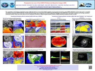
A Phantom for use in an MR Imager. BME 400 October 14, 2005. Team Members: Missy Haehn (Team Leader) Can Pi (BSAC) Ben Sprague (Communications) Andrea Zelisko (BWIG) Advisor: Professor Kristyn Masters Client: Dr. Victor Haughton, M.D. Medical Background.

E N D
A Phantom for use in an MR Imager BME 400 October 14, 2005
Team Members: • Missy Haehn (Team Leader) • Can Pi (BSAC) • Ben Sprague (Communications) • Andrea Zelisko (BWIG) Advisor: • Professor Kristyn Masters Client: • Dr. Victor Haughton, M.D.
Medical Background • Spine consists of vertebrae and disks which act as shock cushions • Disks begin to degenerate with age due to reduced blood flow and water content • This causes disk density loss and risk for vertebrae shifting • Patients suffer from back pain, pinched nerves, muscle spasms, bone spurs
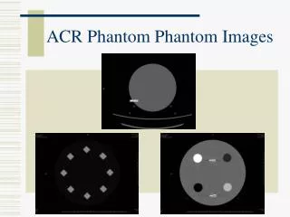
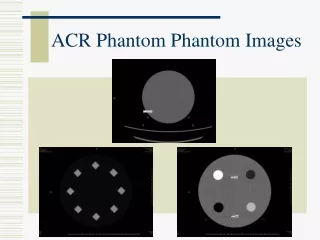
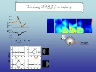
MR Background • MR imaging uses magnets and radio waves to image body • T2 value is relaxation time for protons, relative to water • Can essentially pick a point in the body and ask “What type of tissue are you?” • Phantoms are used to calibrate the scanner as well as in tissue simulations
Motivation • Dr. Haughton is radiologist at • UW-Hospital with a specialty in neuroradiology • Research interest in dynamic spine MR imaging • Specifically disk degeneration • Phantom needed for calibration of laboratory equipment • Mimicking intervertebral disks and T2 values
Client Design Specifications • Hold artificial lumbar disk samples • Contain samples with varying distances to the spinal coil • Include solutions with known relaxation T2 times between 50 and 100 ms • Sit securely atop of the spinal coil • Is easy to use
Current Progress • Current phantoms typically contain water doped solutions • have corresponding T1/T2 values • Constructed by companies such as GE, Supertech, and CIRS • Prices range from $2000 to $5000 • Not directed towards client’s specific research needs
Problems with First Design • Stair structure was large and cumbersome • Material used for structure showed up on MR image • Samples were not close enough together • exposed to varying areas of magnetic field • Disk mimicking samples separated into components over time • Not a leak-proof design
Current Work: Phantom Material • Visited Standard Imaging over summer and received plastic samples • Tissue mimicking • Blue water • Virtual water • Tested samples in MR-found they show up on image and are ideal for x-ray applications • New material to use: Acrylic • Commonly used in commercial and research phantoms
Current Work: Disk Samples • Need artificial disks to maintain integrity over time • Composed of water, collagen, and proteoglycans • Looking into gelatin, agarose, acrylamide, and alginates for substitutes • Tested gelatin and acrylamide in MR with good results • Need to complete testing to decide which is most cost and labor effective
Current Work: Physical Design • Disk samples held close together • Has spigot for easy fill and empty • Able to hold ten samples-two in each tube • Tight seal for phantom-no leaks • Less cumbersome
Potential Problems • Acrylic may show up on MR images • Artifacts from too many interfaces in phantom • Water—plastic—air—glass—sample • Fixed construction does not allow for much variety in experiments • Artificial disk samples may not mimic disks accurately • May need to construct platform so phantom is stable on the MR table/coil
Future Work • Making more hydrogels to test and determining which to use • Continued testing of Gd doped water samples • Construction of phantom • Receiving quote from Acrylix.com • UW polymer processing lab • TESTING!
References • Weidenbaum, M., et al. Correlating Magnetic Resonance Imaging with the Biochemical Content of the Normal Human Intervertebral disk. J. Ortho Research. 10(4): 552-61. • Lumbar Degenerative Disk Disease. DynoMed. 2/12/05. http://www.dynomed.com/encyclopedia/encyclopedia/spine/Lumbar_Degenerative_Disk_Disease.html. • Blechinger, J.C., Madsen, E.L., and Frank, G.R. “Tissue-mimicking gelatin-agar gels for use in magnetic resonance imaging phantoms.” Medical Physics, Vol. 15, No. 4, Jul/Aug 1988. • Phantom Applications and Technology Overview. 2001. Computerized Imaging Reference Systems, Inc. http:www.cirsinc.com/overview.html. • Rice, J. Robin, et all. “Anthropomorphic 1H MRS head phantom.” Medical Physics, Vol. 25, No. 7, July 1998, Part 1. • http://www.supertechx-ray.com/#MRI • http://www.cirsinc.com/productlist.html
Thanks too… • Dr. Victor Haughton • John Perry • Dan Schmidt, Standard Imaging • Professor Bill Murphy • Professor Wally Block • Advisor Kristyn Masters • Ernie Madsen and Maritza Hobson, Medical Physics