Download
1 / 11
120 likes | 435 Views
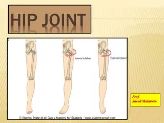
Pathomechanics of hip Joint (part II) practical section. Lecturer: Dr. Manal Radwan Salim Demonstrators: Dr.Mohammed Arafaat Dr. Haytham Essawy Dr. Atef Mohammed Dr. Mai Tolba 6 h practical section Fall 2013-2014 16-11-2013.

E N D
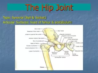
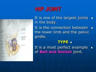
Pathomechanics of hip Joint (part II) practical section Lecturer: Dr. ManalRadwanSalim Demonstrators: Dr.MohammedArafaat Dr. HaythamEssawy Dr. Atef Mohammed Dr. Mai Tolba 6h practical section Fall 2013-2014 16-11-2013
Assessment of joint alignment using motion analysis: 1- identify bony land marks that marks the beginning and ends of each bony segments (forming the tested joint and marks them by colored markers. 2-Fotograph patient in the desired functional position with marks on bony segments ends. 3- Using AutoCAD program draw a straight line that connect between bony segment ends marks. This line represents the anatomical axis of the bony segment .
4- Measure the angle between the two drawn anatomical axes from outside in frontal plane, from anterior in sagital plane for easiness of interpretation at undergraduate level>.5- normal alignment to any joint is calaculted as the antomical position of that joint, for example 0 dorsi flexion of ankle makes 90 degree in it the foot is prependicular to tibia.
Measurements in sagital view 1- measurements of Pelvic Inclination both by using: 1-motion analysis From sagital view if measuring line connecting between so that the line connecting the anterior superior iliac spine and the posterior iliac spine is horizontal. So The pelvis is leveled on hip without anterior or posterior tilt i.e. normal pelvic inclination (50-60 degrees anterior tilt).
4- Pelvis abnormalities: a) Anterior/posterior pelvic tilt: The angle made by a line through the ASIS and the PSIS and the horizontal increases/decreases from an angle of approximately 10-15 degrees. From sagital view
Measurement of Angle of Inclination in x- ray: It the angle between two lines. 1st Method:*line parallel to the ground *line drawn in line with the superior surface of the first sacral vertebrae to the symphysis pubis. 2nd Method:*line parallel to the ground *Line from posterior superior iliac spine to symphysis pubis.
In frontal view c) Pelvic obliquity: A symmetrical height of the pelvis as measured by the iliac crest (posterior view), ASIS in anterior view. in the frontal plane
How to assess knee joint alignment in frontal plane IN OPEN KINEMATIC CHAIN ALL HIP RANGES ARE MEASURED IN SAGITAL AND FRONTAL VIEWS BY ANGLE BETWEEN VERTICAL AND THIGH SEGMENT WHETHER MECHANICAL OR ANATOMICAL AS PREVIOUSLY DISCUSSED AT KNEE
IN closed kinematic chain as the pelvis is moving on femur whether ipsilaterallumbopelvic rhythm in it the lumbar spine moves in the same direction as the the pelvis. For example ( when stopping down ward). Or in contralateralpelvic rhythm where the lumbar spine moves in opposite direction to the pelvic movements as in walking and dancing. When we measure hip ROM we measure the thigh axis to the pelvis axis not to the whole trunk (not as you were taught last year)
In frontal plane we measure the angle between two lines Line connecting two ASIS and the anatomical or mechanical axis of thigh. In sagital view: We measure the angle between thigh axis , and the line extending between greater trochanter and mid point in sagital view along the iliac edge between ASIS and PSIS.
The previous procedures are done in standing on one limb (resembling mid stance) • Initial contact • Loading response • And report to me what are the changes that occurred • These data has no uniform normal yet