Download
1 / 14
230 likes | 541 Views
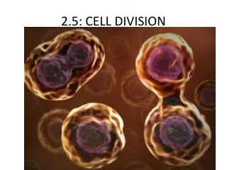
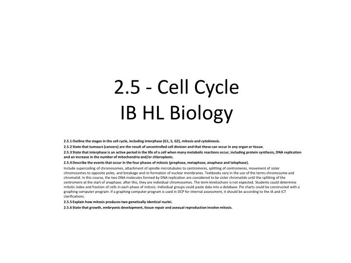
2.5 - Cell Cycle IB HL Biology. 2.5.1 Outline the stages in the cell cycle, including interphase (G1, S, G2), mitosis and cytokinesis. 2.5.2 State that tumours (cancers) are the result of uncontrolled cell division and that these can occur in any organ or tissue.

E N D
2.5 - Cell CycleIB HL Biology 2.5.1 Outline the stages in the cell cycle, including interphase (G1, S, G2), mitosis and cytokinesis. 2.5.2 State that tumours (cancers) are the result of uncontrolled cell division and that these can occur in any organ or tissue. 2.5.3 State that interphase is an active period in the life of a cell when many metabolic reactions occur, including protein synthesis, DNA replication and an increase in the number of mitochondria and/or chloroplasts. 2.5.4 Describe the events that occur in the four phases of mitosis (prophase, metaphase, anaphase and telophase). Include supercoiling of chromosomes, attachment of spindle microtubules to centromeres, splitting of centromeres, movement of sister chromosomes to opposite poles, and breakage and re-formation of nuclear membranes. Textbooks vary in the use of the terms chromosome and chromatid. In this course, the two DNA molecules formed by DNA replication are considered to be sister chromatids until the splitting of the centromere at the start of anaphase; after this, they are individual chromosomes. The term kinetochore is not expected. Students could determine mitotic index and fraction of cells in each phase of mitosis. Individual groups could paste data into a database. Pie charts could be constructed with a graphing computer program. If a graphing computer program is used in DCP for internal assessment, it should be according to the IA and ICT clarifications. 2.5.5 Explain how mitosis produces two genetically identical nuclei. 2.5.6 State that growth, embryonic development, tissue repair and asexual reproduction involve mitosis.
Cell Cycle The Cell Cycle • sequence of events that starts from the time a cell is first formed from a dividing parent cell until its own division into two cells • consists of: • Interphase • Mitotic Phase • mitosiscytokinesis Mitotic Phase
Cell Cycle – Interphase Interphase • accounts for ~ 90% of cell cycle • An active period in the life of a cell when many metabolic reactions occur, such as: • protein synthesis • DNA replication • an increase in the number of mitochondria and/or chloroplasts G1 phase: Cells grow in size S phase: DNA replication occurs (cell still grows) G2 phase: Preparation for cell division (cell still grows)
Cell Cycle – Interphase S Phase Interphase – S Phase
Cell Cycle – Mitotic Phase Mitotic Phase • accounts for ~ 10% of cell cycle • consists of: Mitosis • nucleus and its contents (ie: chromosomes) divide and are evenly distributed to two identical nuclei • unique to eukaryotes • required for: • growth • embryonic development • Replace damaged cells and/or tissue repair • asexual reproduction Cytokinesis • cytoplasm divides in two, creates 2 new identical cells
Mitotic Spindle • Spindle = centrosomes + microtubules + asters • Microtubules: made of subunits of the protein tubulin • While the spindle microtubules assembles, the other microtubules of the cytoskeleton disassemble • Centrosomes are where microtubules assemble • Animal cells contain 2 centrioles at the center of the centrosome, Plant cells do not have centrioles • 1 centrosome replicates during interphase form 2 centrosomes near the nucleus • The duplicated centrosomes move apart during prophase as microtubules grow out from them • At the beginning of metaphase the 2 centrosomes are at opposite ends of the cell • Asters: short microtubules that extends from centrosomes • asters grow until they are in contact with the plasma membrane, by metaphase
Mitosis – Prophase Prophase • the chromatin fibers become supercoiled, condensing into chromosomes • the duplicated chromosome appears as two identical sister chromatids joined together at the centromere • spindle begins to form – composed of centrosomes and microtubules that extend from them. • A pair of centrosomes separate and move towards opposite poles • Animal cells contain centrioles within the centrosomes • nuclear membrane begins to disappear
Mitosis –Metaphase Metaphase • nuclear membrane has disappeared • centrioles have moved to opposite poles of the cell • chromosomes have moved to the equator of the cell • the sister chromatids of every chromosome are attached to the spindle microtubules on opposite poles • the centromeres begin to divide
Mitosis – Anaphase Anaphase • the centromeres have split • the spindle microtubules pull the 2 sister chromatids of a chromosome apart to form 2 chromosomes • each chromatid is now considered a chromosome • the 2 chromosomes begin moving toward opposite ends of the cell, as their microtubules shorten • by the end of the anaphase, the two ends of the cell have equivalent number of chromosomes
Mitosis –Telophase Telophase • the 2 sets of chromosomes have reached the opposite poles of the cell • a nuclear membrane reappear around each set of chromosomes • chromosomes uncoil and become less and less visible • spindle microtubules break down
Cytokinesis • Cytokinesis - the division of the cytoplasm occurs to make 2 daughter cells • Animal cells • cleavage furrows form • the cell is pinched in at the cleavage furrows until 2 cells are formed • Plant cells • a cell plate forms at the equator of cell • a cell wall and cell membranes replace the cell plate • 2 cells are formed
Animal vs Plant Cell Division • Plant Cells • no centrioles • a cell plate forms during cytokinesis was is later replaced by cell membranes & cell wall to divide 1 cell into 2 • Animal Cells • centrioles are found at both poles of cell • Cleavage furrows form at the equator of cell and cause cell to be pinched in until 2 cells are formed
Animal vs Plant Cell Division • Tumours • result of uncontrolled cell division • can grow in any organ • can grow to a large size and can spread to other parts of the body • cancer = diseases caused by the uncontrolled growth of tumours