Download
1 / 28
380 likes | 922 Views
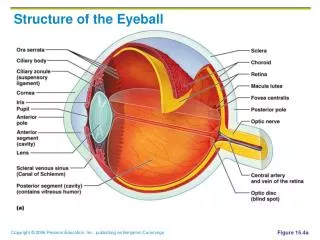
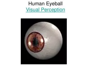
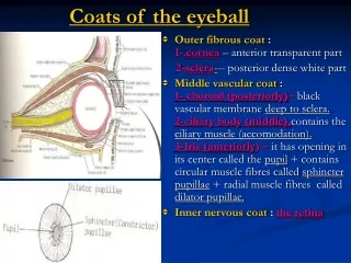
Coats of the eyeball. Outer fibrous coat : 1- .cornea – anterior transparent part 2- sclera - -- posterior dense white part

E N D
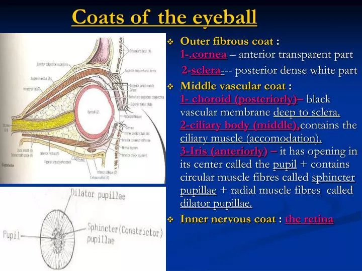
Coats of the eyeball • Outer fibrous coat : 1-.cornea – anterior transparent part 2-sclera--- posterior dense white part • Middle vascular coat :1- choroid (posteriorly)– black vascular membrane deep to sclera.2-ciliary body (middle),contains the ciliary muscle (accomodation).3-Iris (anteriorly) – it has opening in its center called the pupil + contains circular muscle fibres called sphincter pupillae + radial muscle fibres called dilator pupillae. • Inner nervous coat :the retina
The Conjunctiva • It is a thin mucous membrane that lines the inner surface ofeyelids and reflected superiorly & inferiorly onto anterior surface of eyeball to form superior & inferior fornices. • Its epithelium is continuous with that of cornea. • The upper lateral part of superior fornix is pierced by ducts of lacrimal gland.
Levator palpebrae superioris • Origin:roof of orbitabove optic canal , • Insertion:1-by superior lamella (striated muscle) ---anterior surface ofsup. tarsal plate +skin ofupper lid.2-by inferior lamella (smooth m.F)....Upper margin ofsup.tarsal plate. • Nerve :1-striated ms. by oculomotor N.2-smooth ms.by superior cervicalsympathetic F. • Action: elevation of upper eyelid. • Lesion of occulomotor or cervical symp.trunk leads to dropping of upper lid(Ptosis). Sagittal section of eyeball • Tarsal plates: Dense fibrous tissue, lying in the eyelids and containing tarsal glands.
The eyelids • The aponeurosis of insertion of levator palpebraesuperioris pierces the orbital septum to reach superior tarsal plate & skin of upper eyelid. • The superficial surface of tarsal plates + orbital septum are covered by palpebral part of orbicularis oculi muscle. • Action : 1-closed by orbicularis oculi. 2-opened by levator P.S.
The lacrimal apparatus • Lacrimal gland :1-main part (orbital part) lies in lacrimal fossa in anterolateral part of roof of orbit. 2-palpebral part lies in the lateral part of upper eyelid. • It gives 12 ducts : open into superior fornix ofconjunctiva carrying tears to conj.sac.– lacrimal pucti - 2 canaliculi– lacrimal sac (lies in lacrimal groove behind medial palpebral ligament)– nasolacrimal duct– inferior meatus.
The Lacrimal gland • N.supply :Parasymp.secretomotor fibers via lacrimalN. • Preganglionic fibers pass via N. of pterygoid canal to the pterygopalatine ganglion. • Postganglionic fibers leave ganglion via lacrimal N. into lacrimal gland. N.of pterygoid canal
Fascial sheath of the eyeball • It surrounds the eyeball posteriorly from the optic nerve to the corneoscleral junction. • It separates the eyeball from the orbital fat and provides the eyeball with a socket for free movement. • Posteriorly, it fuses with the dural sheath of the optic nerve. • It is pierced by : 1-ciliary nerves & vessels. 2-tendons of orbital muscles , fascial sheath is reflected onto each ms. as a tubular sheaths.
The check & suspensoryligaments of the eyeball. • The fascial sheaths for the tendons of the medial & lateralrecti are attached to the med.& lat. walls of the orbit by medial &lateral check ligaments. • The lower part of fascial sheath below the eyeball is thickened to form suspensory ligament of eye, and connects the check ligaments, so it serves to suspend the eyeball.
The Orbit • What is the common tendinous ring ? • What are the structures passing through the openings in the orbit ? • Muscles of the orbit :1-muscles of eyelids (levator palpebrae superioris). 2-extrinsic muscles of eyeball. 3-intrinsic muscles of eyeball
The Common tendinous ring • It is a fibrous ring of thickening of the periosteum. • It surrounds the optic canal and bridgesthe superior orbital fissure. • It gives origin to the 4 recti muscles. • The sup.R .arises from the upper part of the ring , The inf. R. arises from the lower part of the ring , The med.R. arises from the med.part of the ring. The lat.R. arises by 2-heads from the lateral part of the ring.
What are the structures passing through the openings in the orbit ? Maxillary N.+ inf. ophthalmic V..
The 4-Recti muscles • Origin: common tendinous ring. • Insertion: they form a muscular conethat encloses the optic nerveand post.part of eyeball,/ each tendon of the muscles pierces the fascial sheath of the eyeball and inserted into sclera, 6 mm behind margin of cornea (posterior to corneoscleral junction) • N.supply: sup.,inf.,& medial recti --- oculomotor N./ lateral rectus--- abducent N. • Action:each ms.directs the cornea 1-Superior R.--- upward & medially. 2-Inferior R.--- downward & medially. 3-Medial R.– directs cornea medially 4-lateral R.---laterally.
The 2 oblique Muscles • Superior O.:origin– post.wall of orbital cavity (body of sphenoid). Insertion--- its tendon passes through trochlea (it is afibro-cartilagenous pully attached to frontal bone), then inserted into sup. surface of eyeball (sclera) beneath sup.rectus.N.supply--- trochlear N. Action– rotates eyeball,so that cornea looks downward and laterally. • Inferior O.:origin– floor of orbital cavity. Insertion– lateral surface of eyeball(sclera) ,deep to lateral rectus.N.supply--- N.to inferior oblique from oculomotor N. Action--- rotates eyeball so that cornea looks upwards & laterally.
Intrinsic muscles of eyeball (smooth muscles): • Smooth ,involuntary muscles of circular F (sphincter pupillae) & radiating F. (dilator pupillae)....lying in the iris. • Sphincter PupillaeN.supply : by parasymp.F. from oculomotor N. via N.to inferior oblique after relay in ciliary ganglion, postganglionic fibres pass to eyeball via short ciliary nerves. Action :constricts pupil in bright light & during accommodation. • Dilator Pupillae :N.supply :by symp.F, which pass toeyeball via long ciliary Ns. Action :dilates pupil in low intensity light & in excessive symp. activity as in fright. • Ciliary muscle– smooth muscle in the ciliary body. N.supply– parasymp.F. of 3rdN. as sphincter pupillae muscle.Action : accomodation by making lens more biconvex and increasing the refractive power of lens.
Sensory Nerves :1-optic 2-(lacrimal)3-frontal. 4-(nasociliary) • Optic Nerve :It is the sensory nerve of vision. • Arise from the retina,and peirces the posterior surface of sclera. • It passes through the optic canal ,accompanied by theophthalmic artery (below & lateral to N.) into the middle cranial fossa, where it joins the optic chiasma. • It is surrounded by ciliary nerves& vessels and 4 recti muscles. It is surrounded also by meninges, which fuse with the posterior part of sclera.
Lacrimal N :+parasymp. • It arises from ophthalmic division of trigeminal N. in lateral wall of cavernous sinus. • It enters orbit through S.O.F, outside tendinous ring. • It passes forward above lateral rectus muscle to enter lacrimal gland (parasymp.secretomotor N.). • It ends by supplying skin of lateral part of upper lid. • Frontal N. : • As above in orgin and enters orbit through S.O.F, outside tendinous ring. • It passes forward above L.P.S muscle and ends by dividing into supraorbital & supratrochlear nerves to supply skin of forehead.
Origin : from ophthalmic N. of trigeminal N. in the lateral wall of cavernous sinus. • It enters orbit through sup. Orbital fissure inside the tendinous ring. • It passes medially, crossing above optic N. with ophthalmic artery to reach medial wall of orbit., along upper margin of medial rectus to end bydividing into 2 terminal branches. • Branches :1-sensory root to ciliary ganglion : to supply eyeball through short ciliary Ns. 2-2 long ciliary Ns. : contains sympathetic Fs. To dilator pupillae. 3-posterior ethmoidal N. : to supply ethmoid air sinuses. 4-anterior ethmoidal N. : one of the terminal branches. 5- infratrochlear N. : its 2nd terminal branch. Nasociliary Nerve
Motor nerves:1-Oculomotor 2-Trochlear 3-Abducent • Oculomotor N.: • Sup.division leaves lat.wall of cavernus sinus and enters the orbit through sup.orbital fissure,inside tendinous ring.It supplies sup.rectus & levator P.S. • Inf.division--- as above ,and supplies inf.rectus ,med.rectus ,and inf.oblique.The nerve toinf.oblique gives off preganglionic Fs. to ciliary ganglion and carries parasympathetic fibers to the sphincter pupillae & ciliary muscles, via short ciliary nerves
Trochlear & abducent nerves • Trochlear N. leaves lat.wall of cavernus S. To enter orbit through S.O.F, outside tendinous ring ---- to supply sup.oblique muscle.(SO4). • Abducent N. leaves cavernus sinus to enter orbit through S.O.F, within tendinous ring---- to supply lateral rectus (LR6)
The ciliary ganglion • It is parasympathetic ganglion, situated in theposterior part of the orbitlateral to optic nerve. • It receives preganglionic parasymp.f. from nerve to inf.oblique of oculomotor N. • It sends postganglionic Fs. via short ciliary nerves to the eyeball to supply--- sphincter pupillae &ciliary muscle. • Few sympathetic Fs.pass from internal carotid plexus into orbit without relay in the ganglion.
Ophthalmic artery • It is a branch of the int.carotidartery after it emerges from the cavernous sinus. • It passes through the optic canal below & lateral to the optic nerve, then crosses above it to reach medial wallof orbit. It is now gives off numerous branches. • Branches : ….
Branches of Ophthalmic artery • Centeral artery of retina :itpierces meningeal sheaths of optic N. to enter inside substance of optic N. to reach eyeball (optic disc) and divides into terminal end arteries. • Muscular branches. • Ciliary arteries : anterior group which enter eye at corneoscleral junction & posterior group, which enters near optic N. • Lacrimal artery :to lac.gland. • Supratrochlear & supraorbital arteries to skin of forehead.
Ophthalmic veins • Sup.oph.V.communicates in front with the facial V. • Inf.oph.V.communicates throuth the inf.orbital fissure with the pterygoid venous plexus. • Both veins pass backward through sup.orbital fissure to drain into the cavernous sinus. • No lymph vessels or nodes are present in orbital cavity.
Medial rectus. • Lateral rectus. • Superior rectus. • Inferior rectus. • Superior oblique. • Inferior oblique.
The optic disc • It is the site where the optic N. leaves the retina. • It is the site where it is pierced by the centeralartery of the retina ,and there is complete absence of rods & cones, so that it is insensitive to light and is referred to as * Blind Spot.