Download
1 / 30
300 likes | 413 Views
Anatomy & Physiology. Lymphatic System Ch. 20. Overview of The Lymphatic System. A. Importance of the lymphatic system: 1. Two most importance functions—maintain fluid balance in the internal environment & immunity

E N D
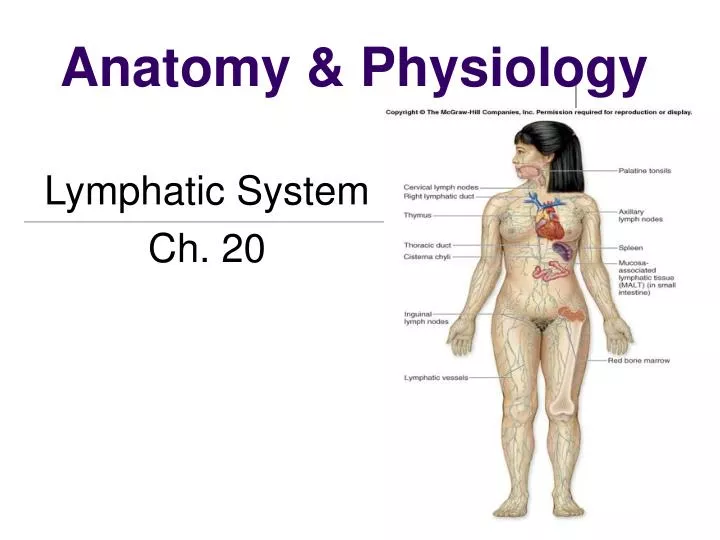
Anatomy & Physiology Lymphatic System Ch. 20
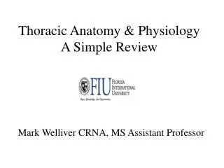
Overview of The Lymphatic System A. Importance of the lymphatic system: 1. Two most importance functions—maintain fluid balance in the internal environment & immunity 2. Lymph vessels act as “drains” to collect excess tissue fluid & return it to the venous blood just before it returns to the heart. 3. Lymphatic System—specialized component of the circulatory system; is made up of: • Lymph • Lymphatic Vessels • Lymph nodes • Isolated nodules of lymphatic tissue • Tonsils • Thymus • Spleen
You’ll need to be able to ID these Lymphatic System organs for your TEST!
Lymph & Interstitial Fluid A. Lymph- 1. Clear, water-appearing fluid found in the lymphatic vessels; closely resembles blood plasma in composition but has a lower percentage of protein; isotonic. 2. Elevated protein concentration in thoracic duct lymph due to protein-rich lymph from the liver and small intestine. B. Interstitial fluid- 1. Complex, organized fluid that fills the spaces between the cells; resembles blood plasma in composition with a lower percentage of protein. 2. Along with blood plasma, constitutes the extra cellular fluid.
Lymphatic Vessels A. Distribution of lymphatic vessels- 1. Lymphatic capillaries-microscopic blind-end vessels where lymphatic vessels originate, wall consists of a single layer of flattened endothelial cells; networks branch and *anastomose freely.*Anastomosis is the connection of two structures. It refers to connections between blood vessels. 2. Lymphatic capillaries merge to form larger lymphatic and eventually form the main lymphatic trunks the right lymphatic ducts and the thoracic duct. 3. Lymph from upper right quadrant empties into right lymphatic duct & then into right subclavian vein. 4. Lymph from rest of the body empties into the thoracic duct, which then drains into the left subclavian vein.
Cont of Lymphatic Vessels… B. Structure of lymphatic vessels- 1. Similar to veins except lymphatic vessels have thinner walls, have more valves and contain lymph nodes. 2. Lymphatic capillary wall is formed by a single layer of thin, flat endothelial cells C. Functions of the lymphatic vessels- 1. Remove high-molecular-weight substances and even particular matter from interstitial spaces. 2. Lacteals absorb fats & other nutrients from the small intestine.
Circulation of Lymph A. The Lymphatic pump 1. Lymph moves through the system in the right direction due to the large number of valves. 2. Breathing movements & skeletal muscle contractions establish a lymph pressure gradient, as they do with venous blood. 3. Lymphokinetic actions—activities that result in a central flow of lymph.
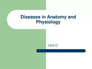
Lymph Nodes A. Structure of Lymph nodes 1. Lymph nodes are oval-shaped structures enclosed by a fibrous capsule. 2. Nodes are similar to biological filter 3. Once lymph enters a node, it moves slowly through sinuses to drain in to efferent exit vessel B. Location of Lymph nodes 1. Most lymph nodes occur in groups 2. Location of groups with greatest clinical importance are submental & submaxillary groups & superficial cervical, superficial cubital, axillary, and inguinal lymph nodes 3. Preauricular lymphs nodes located in front of the ear drain superficial tissues and skin on the lateral side of the head and face.
THE LYMPH NODE -needle like threads spongy bone that surround a network of spaces
Cont. of Lymph Nodes C. Functions of lymph nodes—perform two distinct functions 1. Defense functions: filtration & phagocytosis— reticuloendothelial cells remove microorganisms and other injurious particles from lymph and phagocytose them; if overwhelmed, the lymph nodes can become infected or damaged. 2. Hematopoiesis—process of blood cell formation, lymphatic tissue is the site for the final stages of maturation of some lymphocytes & monocytes.
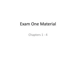
Lymphatic drainage of the breast A. Distribution of lymphatics in the breast 1. Drained by two sets of lymphatic vessels a. Lymphatics that drain the skin over the breast with the exception of the areola & nipple b. Lymphatics that drain the substance of the breast as well as the skin of the areola & nipple 2. Superficial vessels coverage to form a diffuse, cutaneous lymphatic plexus 3. Subareolar plexus-this is located under the areola surrounding the nipple, where communication between the cutaneous plexus & larger lymphatics that drain the secretory tissue & ducts of the breast occurs
Pectoralis major muscle Axillary lymph nodes: Levels I Axillary lymph nodes: Levels II Axillary lymph nodes: Levels III Subclavicular lymph nodes Internal mammary nodes
Tonsils A. Location- under the mucous membranes in the mouth & back of the throat. 1. Palatine tonsils- located on each side of the throat. 2. Pharyngeal tonsils- located near the posterior opening of the nasal cavity. 3. Lingual tonsils- located near the base of the tongue B. Function- Protect against bacteria that may invade tissues around the openings the nasal & oral cavities.
Thymus A. Location & appearance of the Thymus 1. Primary central organ of lymphatic system 2. Single, unpaired organ located in the mediastinum, extending upward to the lower edge of the thyroid & inferiorly as far as the 4th costal cartilage 3. Thymus is pinkish gray in children and wish advancing age, becomes yellowish as lymphatic tissue is replaced by fat.
B. Structure of the Thymus 1. Pryamid-shaped lobes are subdivided into small lobules
Cont. of Structure of the Thymus 2. Each lobule is composed of a dense cellular cortex & an inner, less dense, medulla 3. Medullary tissue can be identified by presence of Thymic corpuscle.
C. Function of the Thymus 1. Plays vital role in immunity mechanism 2. Source of lymphocytes before birth 3. Shortly after birth, the thymus secretes Thymosin, which enables lymphocytes to develop into T-Cells….
Spleen A. Location-in the left hypochondrium (either one of two regions of the abdomen), directly below the diaphragm, above the left kidney & descending colon, & behind the fundus (the base of an organ) of the stomach!
Cont of Spleen B. Structure of the Spleen 1. Ovoid in shape 2. Surrounded by fibrous capsule with inward extensions that divide the organ into compartments. 3. White pulp-dense masses of developing lymphocytes 4. Red pulp-near outer regions, made up of a network of fine reticular fibers submerged in blood that comes from nearby arterioles. C. Functions of the Spleen 1. Defense-macrophages lining the sunusoids of the spleen remove microorganisms from the blood & phagocytose them 2. Hematopoiesis-monocytes & lymphocytes complete their development in the spleen. 3. Red blood cell and platelet destruction-macrophages remove worn-out RBC’s and imperfect platelets and destroy them by phagocytosis; also salvage iron and globin from destroyed RBC’s
Don’t confuse Lymphedema with Edema… We’ll get to this later!
Cycle of Life: Lymphatic System A. Dramatic changes throughout life B. Organs with lymphocytes appear before birth and grow until puberty C. Postpuberty 1. Organs atrophy (shrink in size, degenerate) through late adulthood and become fatty or fibrous. 2. Spleen-it develops early and remains intact
The Big Picture:The Lymphatic System & The Whole Body A. Lymphatic system drains away excess water from large areas B. Lymph is conducted through lymphatic vessels to nodes, where contaminants are removed. C. Lymphatic system benefits the whole body by maintaining fluid balance and freedom from disease
What Is Lymphedema? Lymphedema is an accumulation of lymphatic fluid in the interstitial tissue that causes swelling, mostly in the arm(s) and/or leg(s), and occasionally in other parts of the body. Lymphedema can develop when lymphatic vessels are missing or impaired (primary), or when lymph vessels are damaged or lymph nodes removed (secondary). When the impairment becomes so great that the lymphatic fluid exceeds the lymphatic transport capacity, an abnormal amount of protein-rich fluid collects in the tissues of the affected area. Left untreated, this stagnant, protein-rich fluid not only causes tissue channels to increase in size and number, but also reduces oxygen availability in the transport system, interferes with wound healing, and provides a culture medium for bacteria that can result in lymphangitis (infection). Lymphedema should not be confused with edema resulting from venous insufficiency, which is not lymph-edema. However, untreated venous insufficiency can progress into a combined venous/lymphatic disorder which is treated in the same way as lymphedema.
What Causes Lymphedema? Primary lymphedema, which can affect from one to as many as four limbs and/or other parts of the body, can be present at birth, develop at the onset of puberty (praecox) or in adulthood (tarda), all from unknown causes, or associated with vascular anomolies such as: Port Wine Stain, and Klippel Trenaury (a rarecongenital medical condition in which blood vessels and/or lymph vessels fail to form properly). Secondary lymphedema, or acquired lymphedema, can develop as a result of surgery, radiation, infection or trauma. Specific surgeries, such as surgery for melanoma or breast, head and neck, prostate or testicular, bladder or colon cancer, all of which currently require removal of lymph nodes, put patients at risk of developing secondary lymphedema. If lymph nodes are removed, there is always a risk of developing lymphedema. Port Wine Stain
Virus Project…..due May 4th! If you have any questions about your projects, please see me or email! Please email your Virus Project powerpoints: Paula.Carrasco@humble.k12.tx.us
STUDY & Prepare for your TEST • Study the Immune System & Lymphatic System powerpoints for your TEST!!! • Your TEST will be Wednesday, MAY 4th. DON’T LOOSE FOCUS!! STUDY NOW MORE THAN EVER!!!!!!