Download
1 / 10
100 likes | 394 Views
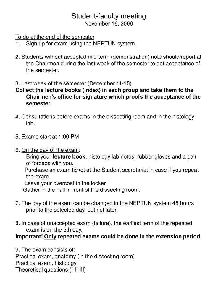
Student-faculty meeting November 16, 2006 To do at the end of the semester Sign up for exam using the NEPTUN system. 2. Students without accepted mid-term (demonstration) note should report at the Chairmen during the last week of the semester to get acceptance of the semester.

E N D
Student-faculty meeting • November 16, 2006 • To do at the end of the semester • Sign up for exam using the NEPTUN system. • 2. Students without accepted mid-term (demonstration) note should report at the Chairmen during the last week of the semester to get acceptance of the semester. • 3. Last week of the semester (December 11-15). • Collect the lecture books (index) in each group and take them to the Chairmen’s office for signature which proofs the acceptance of the semester. • 4. Consultations before exams in the dissecting room and in the histology lab. • 5. Exams start at 1:00 PM • 6. On the day of the exam: • Bring your lecture book, histology lab notes, rubber gloves and a pair of forceps with you. • Purchase an exam ticket at the Student secretariat in case if you repeat the exam. • Leave your overcoat in the locker. • Gather in the hall in front of the dissecting room. • 7. The day of the exam can be changed in the NEPTUN system 48 hours prior to the selected day, but not later. • 8. In case of unaccepted exam (failure), the earliest term of the repeated exam is on the 5th day. • Important! Only repeated exams could be done in the extension period. • 9. The exam consists of: • Practical exam, anatomy (in the dissecting room) • Practical exam, histology • Theoretical questions (I-II-III)
Topics of the end-of-semester exam (colloquium) for medical and dental students Session 2006/2007 First semester Practical exam, anatomy (in the dissecting room) Demonstration on preparations Skull Anterior cranial fossa (composition, boundaries, connections) Middle cranial fossa (composition, boundaries, connections) Posterior cranial fossa (composition, boundaries, connections) Walls and connections of the orbit Walls and connections of the nasal cavity Inferior surface and connections of the base of the skull Bony walls of the oral cavity, the temporal and infratemporal fossa Walls and connections of the pterygopalatine fossa Joints of the extremities Muscles, vessels and nerves of the extremities (without the cutaneous nerves) Practical exam, histology Study of a histological specimenConcept of basic tissues Definition and classification of epithelial tissue Simple epithelia Stratified epithelia Membrane specializations of epithelia Glandular epithelia Pigment epithelium and sensory epithelium Cells of connective tissue Ground substance and fibers of connective tissue Types of connective tissue Umbilical cord and placenta Blood and the formed elements of blood Histology of the bone marrow and the maturation of erythrocytes and platelets Differentiation of granulocytes, lymphocytes and monocytes Histology of cartilage Histology of the bone Intramembranous ossification Endochondral ossification Growth and remodeling of bone Smooth muscle and myoepithelial cells Histology of the skeletal muscle Histology of the cardiac muscle Histology of the neuron Histology of the neuroglia Supporting cells in the peripheral nervous system Nerve fibers, myelin sheath Histology of the sensory receptors Effectors: motor end-plate and grundplexus Interneuronal synapses
Theoretical exam, first question Description of a joint (surfaces, capsule, ligaments, type, axes, movements) and the muscles acting on the joint Fibrous and cartilaginous joints Components of the synovial joints Classification of synovial joints; movements and mechanisms Structure of the vertebral column and the gross anatomy of the muscles responsible for its movements Movements of the head (atlantooccipital and atlantoaxial joints) and the gross anatomy of the muscles participating in them Joints of the shoulder girdle and the gross anatomy of the muscles acting on them The shoulder joint and the gross anatomy of the muscles acting on it The elbow joint and the gross anatomy of the muscles acting on it Structure and movements at the wrist joint (radiocarpal joint) and the gross anatomy of the muscles acting on it Joints of fingers (metacarpophalageal and interphalangeal joints) and the gross anatomy of the muscles concerned in their movements Joints of the thumb (carpometacarpal, metacarpophalangeal and interphalangeal joints of the thumb) and the gross anatomy of the muscles concerned in their movements The hip joint and the gross anatomy of the muscles concerned in its movements The knee joint and the gross anatomy of the muscles concerned in its movements Ankle joint and the gross anatomy of the muscles concerned in its movements Subtalar and talocalcaneonavicular joints and the gross anatomy of the muscles acting on them Temporomandibular joint and the gross anatomy of the muscles acting on it
Theoretical exam, second question Topic in the locomotor system unrelated to joints Architecture and classification of bones Structure and actions of somatic muscles Osteofibrous structure of the thoracic cage (bones, joints, ligaments, movements) Muscles and movements of the thorax Muscles of the back and nape (occipital region) The axilla, the quadrangular and triangular spaces The cubital fossa Muscles and cross section of the arm Muscles and cross section of forearm Osteofibrous spaces and muscle compartments of the hand, tendon sheaths Structure of the osteofibrous pelvis (bones, ligaments and membranes) Muscles of the buttock, the posterior abdominal wall and the pelvis (external and internal muscles of the hip) Osteofibrous compartments, muscles and cross section of the thigh Popliteal fossa Femoral sheath, vascular and muscular compartments; adductor canal Osteofibrous compartments, muscles and the cross section of the leg Structure of the foot, arches of the foot Osteofibrous compartments of the foot, tendon sheaths Muscles of mastication Diaphragm Lateral abdominal muscles and fasciae Rectus abdominis muscle and the rectus sheath Inguinal canal Femoral canal Superficial muscles of the neck and the muscle triangles Deep muscles of the neck and the laminae of the cervical fascia Muscles of facial expression
Theoretical exam, third question Embryology Spermatogenesis Oogenesis Fertilization, cleavage of the zygote Blastocyst formation; the bilaminar embryonic disc Implantation Formation of the intraembryonic mesoderm; the notochord Neurulation (neural tube and neural crest) Differentiation of the intraembryonic mesoderm; formation and derivatives of the somites Derivatives of the intermediate mesoderm Lateral plate mesoderm and its derivatives Folding of the embryo Development of the primitive cardiovascular system and the placental circulation The structure and function of the placenta Development of the fetal membranes (chorion and amnion) and the umbilical cord The embryonic and fetal periods Twin formation Development of the limbs Development of the vertebral column Development of the skull Development of the skeletal muscular system
Topics of the end-of-semester exam (colloquium) for medical and dental students Session 2006/2007 Third semester Practical exam, anatomy (in the dissecting room) Demonstration on preparations Gross anatomy of the brain (meninges, arteries, veins, dura mater sinuses, circulation of the cerebrospinal fluid) Regions on the dorsal surface of the trunk and extremities Regio nuchae Regio dorsalis scapulae Regio deltoidea Regio brachii posterior Regio cubiti posterior Regio antebrachii dorsalis Regio carpi dorsalis Regio dorsalis manus Foveola radialis Regio glutea Regio femoris posterior Regio poplitea Regio cruris posterior Regio malleolaris lateralis Regio plantaris Back region
Practical exam, histology Study of a histological specimen Microscopic structure of the nervous tissue and the nervous system Endocrine organs Gross anatomy and histology of the pituitary gland; development of the neural lobe Blood supply, histology and development of the distal lobe of the pituitary gland Gross and microscopic anatomy of the pineal gland Gross and microscopic anatomy as well as development of the thyroid gland Gross and microscopic anatomy as well as development of the parathyroid gland Gross and microscopic anatomy as well as development of the suprarenal gland Histology of the Langerhans islets Endocrine cells and function of the male and female gonads Microscopic structure of the eyeball Microscopic structure of the cochlea Microscopic structure of the skin including the mammary gland
Theoretical exam, first question Microscopic structure and development of the central nervous system Development and primary differentiation of the neural tube Development of the spinal cord; neurohistogenesis Differentiation of the prosencephalon vesicle; development of the hemispheres and the lateral ventricle Differentiation of the diencephalon vesicle, development of the third ventricle Differentiation of the mesencephalon and rhombencephalon vesicles, development of the fourth ventricle Roots, branches and components of the spinal nerves; spinal segment Fine structure (microscopy) of the spinal cord Neurons and function of the spinal proprioceptive (strech) reflex Neurons and function of the spinal flexion (withdrawal) reflex Neurons and function of the visceral reflex Microscopic anatomy of the medulla Microscopic anatomy of the pons Microscopic anatomy of the midbrain Nuclei of the cranial nerves Tracts of the brainstem (medulla, pons, midbrain) Microscopic anatomy of the cerebellum Afferent and efferent connections of cerebellum Microscopic anatomy of thalamus Hypothalamus, hypothalamo-hypophyseal systems Microscopic anatomy of the basal ganglia Histology of the cerebral cortex; cortical fields Internal capsule Tracts of the protopathic sensibility Tracts of the epicritic sensibility Corticospinal tract (pyramidal tract) Extrapyramidal system Limbic system (nuclei and tracts)
Theoretical exam, second question Gross anatomy and development of the peripheral nervous system Development and differentiation of the cells in the neural crest Development of the peripheral nervous system Nuclei and branches of the IIIrd, IVth and VIth cranial nerves Nuclei of the trigeminal nerve; course and fiber composition of the branches of the ophthalmic (V/1) nerve Course and fiber composition of the branches of the maxillary nerve (V/2) Course and fiber composition of the branches of the mandibular nerve (V/3) Nuclei, course and fiber composition of the branches of the facial nerve (VII) Nuclei, course and fiber composition of the branches of the glossopharyngeal nerve (IX) Nuclei, course and fiber composition of the branches of the vagus nerve (X) Nuclei, course and fiber composition of the branches of the accessory (XI) and hypoglossal nerves (XII) Cervical plexus and its branches Brachial plexus and its short branches to the neck and shoulder girdle General structural plan of the autonomic nervous system The sympathetic trunk Cranial part of the parasympathetic nervous system
Theoretical exam, third question Gross anatomy, histology and embryology of the organs of special senses Gross anatomy and microscopic structure of the fibrous coat of the eye ball (cornea, sclera) Gross anatomy and microscopic structure of the vascular coat of the eye ball (choroid, ciliary body, iris) Gross anatomy, microscopic structure and development of the nervous coat of the eye ball (retina) Neurons of the visual pathways; localization and microscopic structure of the visual cortex Gross anatomy, microscopic structure and development of the lens, accomodation Gross anatomy and content of the chambers of the eye, circulation of the aqueous humor; gross anatomy of the vitreous body Gross anatomy and function of the external eye muscles. Visual reflexes Gross anatomy, microscopic structure of the eye lids; conjunctiva, Tennon's capsule and periorbit Gross anatomy, microscopic structure and development of the lacrimal apparatus Gross anatomy and development of the external ear and the tympanic membrane Gross anatomy and development of the tympanic cavity and the auditory tube Gross anatomy and development of the auditory ossicles; joints, muscles, and mucous membrane of the tympanic cavity Sensory innervation and blood supply of the tympanic cavity Gross anatomy of the bony labyrinth Gross anatomy and development of the labyrinth Neuronal structure of the vestibular system Gross anatomy, microscopic structure and development of the cochlear duct and the organ of Corti Neurons of the auditory pathways Internal acoustic meatus Organ and pathways of olfaction Organ and pathways of taste