Download

1 / 1
10 likes | 128 Views
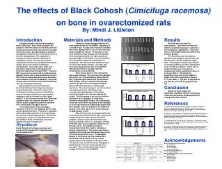
A. B. C. *. *. *. *. *. *. *. *. *. Figure 2 . MCF-7 cells on day 10 of proliferation assay. 10x magnification. Scale bar = 100µm. A - Cimicifuga racemosa at 100% concentration. B - Cimicifuga racemosa at 25% concentration. C - 17b-Estradiol 1x10 -7 M. . *. *. *. *. *. *.

E N D
A B C * * * * * * * * * Figure 2. MCF-7 cells on day 10 of proliferation assay. 10x magnification. Scale bar = 100µm. A - Cimicifuga racemosa at 100% concentration. B - Cimicifuga racemosa at 25% concentration. C - 17b-Estradiol 1x10⁻⁷ M. * * * * * * * * * * * * Investigating the Mechanism of Action of Cimicifuga racemosa Introduction In recent years, hormone replacement therapy (HRT) has come under scrutiny for potential side effects, such as stimulating breast cancer. Traditionally HRT has been used to treat the symptoms of menopause. However, with these concerns about HRT, herbal medicines, such as Black Cohosh (Cimicifuga racemosa) have been introduced as a potentially safe alternative. This study seeks to elucidate the mechanism of action by which black cohosh has its effect. HRT (estrogen treatment) has been used to treat the symptoms of menopause, which include night sweats, hot flashes and mood swings. However, in women with a history of estrogen-dependent tumors, or a family history of breast cancer, HRT is avoided as a link has been established between breast cancer and postmenopausal HRT.2,4,13Cimicifuga racemosa (CR; black cohosh) has been used to relieve these symptoms and appears to be an effective5,13 and safe alternative.4 However, it is hypothesized that CR may contain estrogenic compounds, and alleviate symptoms via the same pathway as HRT. Should this be true, the risks associated with HRT may also be true for CR. Two theories concerning the mechanism of action of CR have been proposed; one, that CR acts via an estrogen receptor (ER), much like HRT, or two, that CR exhibits SERM (selective estrogen receptor modulator) activity.2,5 There are two known estrogen receptors in the human; ER and ER. Jarry et al have demonstrated that a CR extract does not bind to either ER or ER, strongly suggesting that CR does not act via an estrogen receptor.5 However, a third ER, designated ER, has been identified in Atlantic croaker fish.3 As CR appears to exhibit estrogen-like qualities (cessation of hot flashes, night sweats and mood swings), it has been hypothesized that though CR does not act through either ER or ER, it may act via ERto cause the observed estrogenic effects. Alternatively, a SERM compound would both stimulate and inhibit estrogen effects at various concentrations.7 The opposing actions (stimulatory or inhibitory) of these SERM compounds depend on the target tissue (breast, uterus, bone).7 This hypothesis is supported by current research, which has shown CR to inhibit estrogen-dependent cell lines, while still appearing to have an estrogenic effect, as evidenced by the alleviation of hot flashes seen in ovariectomized rats and double-blind placebo-based clinical trials.5,7,11,12 To investigate the mechanism of action of CR, an MCF-7 cell proliferation assay, originally developed by Soto et al, was used.10 Using such an assay, it can be determined if CR is an estrogen mimic by analyzing the cell growth, or proliferation of these estrogen-dependent cells.10 Hypothesis: Cimicifuga racemosa will have an inhibitory effect on MCF-7 cells. Cell growth will be compared to the controls (medium and ethanol). 17ß-Estradiol and Tamoxifen will act as positive and negative controls, respectively. Materials and Methods Cell Culture MCF-7 cells were obtained from the American Cell Culture Type Collection, and cultured in Dulbecco’s Modified Eagles Medium (Sigma-Aldrich) without phenol red and supplemented with 5% fetal calf serum (Sigma-Aldrich), 1% penicillin-streptomycin (Sigma-Aldrich) and 0.584 g/L L-glutamine (Sigma-Aldrich). The cells were cultured in 75 cm2 flasks at 37°C and 5% CO2. These conditions were also used for the proliferation assays. Proliferation Assay For the proliferation assays, fetal calf serum (FCS) was stripped with dextran-coated charcoal. The charcoal stripped fetal calf serum (CS-FCS) was used in all proliferation assays. The CS-FCS was prepared by mixing dextran-coated charcoal (Sigma-Aldrich) with FCS, and incubating the mixture in a water bath at 56C for 2x45 minutes, centrifuged at 3000 rpm for 10 minutes and the resulting suspension sterile filtered through a 0.22m filter (Sigma-Aldrich).2 Cells were plated on a 96-well plate at 10,000 cells per well (200l; 0.1% EtOH) and dosed with 17-estradiol (1x10⁻⁸M).6,8,9 After 48 hours, the medium was removed, and the wells washed with 50µl phosphate buffered saline (PBS). The PBS was removed, and replaced with non-estrogen-containing medium (200µl/well) for an additional 48 hours.9 The cells were then dosed with 17-estradiol (1x10⁻⁷M– 1x10-12M), Tamoxifen (1x10⁻⁶M– 1x10-11M), or Cimicifuga racemosa (100%, 85, 70. 55, 40, and 25%). 17b-Estradiol and Tamoxifen were both used as controls, as 17b-Estradiol is a known stimulator of MCF-7 cells, and Tamoxifen is known to inhibit cell growth. Interestingly, the CR also inhibited cell growth, at all but the lowest concentration, which is similar to what has been observed in previous studies.2 In Figure 2A and B, it is possible to see fewer cells at a 100% CR concentration, versus a 25% concentration. In comparison, Figure 2C shows the inhibitory action of 17b-estradiol (1x10⁻⁷M). There appear to be fewer cells at 25%CR than in the 17b-estradiol, which seems to support the results obtained from the Bradford reaction. While CR did inhibit MCF-7 cell growth at high concentrations (Figure 5), the results are questionable as the positive and negative controls failed to produce the expected results. While this data does support the hypothesis that CR could inhibit MCF-7 cell growth, it does not explain the mechanism of action. It is feasible that CR could still be binding to either ER or ER to cause such an antagonistic effect, much like Tamoxifen, or CR could have an inhibitive effect via another pathway. To examine synergistic activity, Cimicifuga racemosa was tested at a variety of concentrations in either 1x10⁻⁸M 17-estradiol or 1x10⁻⁶M Tamoxifen. The interaction between 17-estradiol and Tamoxifen was also examined, with Tamoxifen concentrations (1x10⁻⁶M– 1x10-11M) tested at a steady 1x10⁻⁸M 17-estradiol concentration. This also served as a control to test the possible inhibitory effects of CR. After 6 days, the medium was removed, and the wells washed with PBS. The cells were then lysed with a standard lysis solution (Tris-HCL, 06057g; 1% Triton X-100; Sodium deoxycholate, 0.2g; SDS, 0.2g; EDTA, 0.2 ml). Per the methods of Bradford et al, the average protein concentrations per well were determined by recording the absorbance at 562nm on a Thermo Spectronic spectrophotometer.1 This gave the average amount of protein per well, not the number of cells per well. The protein levels were used in place of actual cell counts to quantify the growth of the cell cultures. The data were then analyzed using aStudent’s t-test. Preparation of Chemicals The CR extract was prepared from capsules obtained from Vitamin World. Capsules contained 540 mg of CR root material (suggested daily dose) in a gelatin casing. The capsule was broken open and the gelatin casing discarded. The contents of 5 capsules (2.672g) were extracted with 5 volumes of 50% EtOH (13.36 ml) for 24 hours. The CR dilutions (100%, 85, 70. 55, 40, and 25%) were also prepared in 50% EtOH. Both the 17ß-Estradiol and Tamoxifen (Sigma-Aldrich) were prepared as 1x10⁻⁸M and 1x10⁻⁶M stock solutions in 50% EtOH, respectively. These stock solutions were used to form the dilution series used in the proliferation assays. Figure 5. Cimicifuga racemosa extract at various doses. All data shown with mean ± SD; n = 8. *p < 0.05 compared to control (Student’s t-test). It has been hypothesized that CR may act upon the hypothalamus to alleviate hot flashes and night sweats.13 This suggests that CR may be a SERM compound if it mimics estrogen in some organs, yet not others. The MCF-7 cell assay does not provide definitive proof for the estrogenic potential of a compound, yet it can be used effectively to evaluate several compounds at once in one tissue type. A third hypothesis, postulated by Hostanksa et al, evaluated the apoptotic effect of CR upon cell lines.4 CR may stimulate apoptosis in cells, resulting in inhibition. This theory is supported by this study, as the cell plating density did not appear to be a factor for the cells dosed with CR. This suggests that CR may be actively inhibiting cells by such a pathway as apoptosis. Future Experiments As shown in Figure 6, a cell density of 5,000 cells per well (200µl) appears to be the best initial plating density. At 10,000 cells per well there is no significant difference between dosing the cells with 17ß-Estradiol or Tamoxifen. At both 2,000 and 5,000 cells per well however, a significant difference is noted. Therefore, future experiments should investigate the effect of plating various cell densities. Furthermore, an alternative form of quantifying cells should be used. A Coulter counter or a hemocytometer may be more accurate when dealing with small samples of protein, and less susceptible to “noise.” Conclusion Cimicifuga racemosa was shown to inhibit MCF-7 cells at all but the lowest concentration, yet the positive and negative controls were ineffectual, most likely due to the initial cell plating density. This suggests that, as CR did have a consistently inhibitory effect at high concentrations, the extract may be killing MCF-7 cells. Such an occurrence could occur via cytotoxicity, apoptosis, or other undetermined pathways. The exact mechanism of action of CR cannot be determined, and will require further experimentation. Diana Rohlman, Dr. David Brown Marietta College Biology Department Figure 4. Dose response curve of Tamoxifen on MCF-7 cells. Cells dosed at day 6 of proliferation assay. All data shown with mean ± SD; n = 8. *p < 0.05 compared to EtOH control (Student’s t-test). Figure 3. Dose response curve of 17ß-Estradiol on MCF-7 cells. Cells dosed at day 6 of proliferation assay. All data shown with mean ± SD; n = 8. *p < 0.05 compared to EtOH control (Student’s t-test). • Results and Discussion • An MCF-7 cellproliferation assay was used in an attempt to determine the mechanism of action of Cimicifuga racemosa. A modification of the Bradford reaction was used to quantify the cells, using BCA reagents in place of the Bradford reagent. • Initial experimentation with the BCA reagent showed that varying cell densities could be accurately determined (Figure 1). For these experiments, cells were plated at various concentrations (25,000-200,000 cells/well). These concentrations were chosen to reflect the expected cell concentrations after the 10-day proliferation assay. After 24 hours, the cells were lysed, transferred to a cuvette with 1ml BCA reagent and incubated at room temperature for 1 hour. The Bradford reaction was then conducted to quantify protein levels. • In the actual proliferation assays, the positive control (17b-estradiol) did not stimulate MCF-7 cell growth. At higher concentrations, 17b-estradiol actually inhibited cell growth (Figure 3). This was unexpected, as 17b-estradiol has been shown in previous studies to stimulate cell growth at the concentrations used.2Cell plating density was investigated to determine if this was a contributing factor. As seen in Figure 6, at 10,000 cells per well there was no significant difference between 17b-estradiol and Tamoxifen, yet there were significant differences at cell plating densities. 17b-estradiol (1x10⁻⁸M) stimulated cell growth, while Tamoxifen (1x10⁻⁶M) inhibited cell growth at low densities. This suggests that the initial cell plating density was too high, and may have contributed to the unexpected results. • The negative control (Tamoxifen) consistently inhibited cell growth (Figure 4) at higher concentrations. However, this conflicts with previous research that showed a stimulatory effect at 1x10⁻⁷M Tamoxifen.2 Again, the initial cell plating density appears to be a factor in these results. Figure 6.MCF-7 cell plating density. (n = 16: 17ß-Estradiol; n = 8: Tamoxifen) All data shown with mean ± SD. *p < 0.05 17ß-Estradiol compared to Tamoxifen. • Works Cited • 1. Bradford MM. (1976) Anal.Biochem. 72: 248-254. • 2. Bodinet C, Freudenstein J. (2002) Breast Cancer Research & Treatment 76: 1-10. • 3. Hawkins MB, Thornton JW, Crews D, Skipper JK, Dotte A, Thomas P. (2000) Proc. Nat’l Acad. of Sciences USA 97(20): 10751-10756. • 4. Hostanksa K, Nisslein T, Freudenstein J, Reichling J, Saller R. (2004) BreastCancer Research & Treatment 84: 151-160. • 5. Jarry H, Metten M, Spengler B, Christoffel V, Wuttke W. (2003) Maturitas 44(Supplement 1): S31-S38. • 6. Jones PA, Baker VA, Irwin A, Earl LK. (1997) Toxicology in Vitro 11:769-773. • 7. Muñoz GH, Pluchino S. (2003) Maturitas 44(Supplement 1): S59-S65. • 8. Payne J, Jones C, Lakhani S, Kortenkamp A. (2000) The Science of the Total Environment 248: 51-62. • 9. Rasmussen TH, Nielsen JB. (2002) Biomarkers 7(4): 322-336. • 10. Soto AM, Sonnenschein C, Chung KL, Fernandez MF, Olea N, Serrano FO. (1995) Environ. Health Perspect.103(Supplement 7): 113-122. • 11. Vermes G, Bánhidy F, Ács N. (2005) Advances in Therapy 22(2): 148-154. • 12. Winterhoff H, Spengler B, Christoffel V, Butterweck V, Löhning A. (2003) Maturitas 44(Supplement 1): S51-S58. • 13. Wuttke W, Seidlová-Wuttke D, Gorkow C. (2003) Maturitas 44(Supplement 1): S67-S77. Acknowledgements: Dr. David Brown, capstone advisor; Dr. Peter Hogan, capstone class advisor; Dr. Kevin Pate for help in the CR ethanol extraction; 2007 Senior Biology Capstone Class, in particular: Mindi Littleton, Andrea Marion, and Mike Braun. Figure 1. Testing the Bradford Reaction at known concentrations of MCF-7 cells. Cells incubated for 24 hour in a 96-well plate, then lysed and treated with BCA Reagent. Absorbency recorded using a Thermo Spectronic spectrophotometer.