Download
1 / 51
581 likes | 1.74k Views
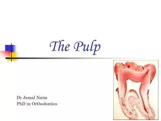
PULP. GENERAL FEATURES Every person normally has a total of 52 pulp organs , 32 in the permanent & 20 in the primary teeth. The total volumes of all the permanent teeth pulp organs is 0.38 cc. The mean volume of a single adult human pulp is 0.02 cc.

E N D
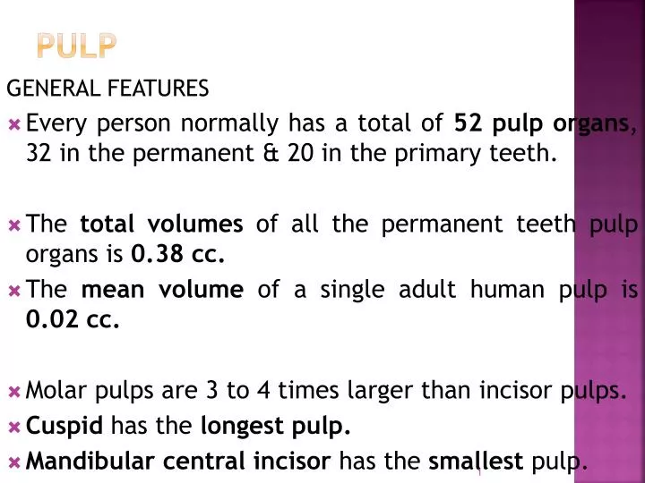
PULP GENERAL FEATURES • Every person normally has a total of 52 pulp organs, 32 in the permanent & 20 in the primary teeth. • The total volumes of all the permanent teeth pulp organs is 0.38 cc. • The mean volume of a single adult human pulp is 0.02 cc. • Molar pulps are 3 to 4 times larger than incisor pulps. • Cuspid has the longest pulp. • Mandibular central incisor has the smallest pulp.
APICAL FORAMEN • The average size of apical foramen of the maxillary teeth in the adult is 0.4 mm. • In the mandibular teeth it is slightly smaller, being smaller, being 0.3 mm in diameter. ACCESSORY CANALS • Found in apical third region & furcation region.
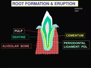
PRIMARY PULP ORGANS • The average length of time a primary pulp functions in the oral cavity is only about 8.3 years. • This amount of time can be divided into three periods :- • Pulp organ growth • Pulp maturation • Pulp regression
PERMANENT PULP ORGANS • Pulp of the permanent teeth undergoes development for about 12 years, 4 months. • The maxillary arches require slightly longer to complete each process of development than do the mandibular arches.
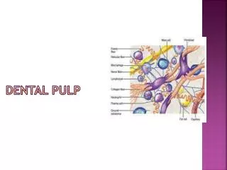
STRUCTURAL FEATURES The pulp is circumscribed by the specialized odontogenic region composed of :- • The odontoblasts (the dentin forming cells) • The cell-free zone (Weil’s zone), and • The cell-rich zone
FIBROBLASTS • Are the most numerous cell types in the pulp. • Function in collagen fiber formation . • Have the typical stellate shape & extensive processes that contact & are joined by intercellular junctions. • Have abundant rough-surfaced endoplasmic reticulum, mitochondria & other cell organelles. • Also have the capability of ingesting & degrading this same matrix.
In the older pulp, they appear rounded or spindle shaped with short processes & exhibit fewer intracellular organelles. They are then termed fibrocytes.
UNDIFFERENTIATED MESENCHYMAL CELLS • They are the primary cells in the very young pulp. • Only a few are seen in the pulps after root completion. • Appear larger than fibroblasts. • Are polyhedral in shape with peripheral processes & large oval nuclei. • Are found along pulp vessels, in the cell-rich zone & scattered throughout the central pulp.
Viewed from the side, they appear spindle shaped. • They are believed to be a totipotent cell & when need arises they may become odontoblasts, fibroblasts, or macrophages. • Decrease in number in old age.
ODONTOBLASTS • The second most prominent cell in the pulp. • Reside adjacent to the predentin with cell bodies in the pulp & cell processes in the dentinal tubules. • About 5 to 7 um in diameter & 25 to 40 um in length. • Cell bodies are columnar in appearance with large oval nuclei, which fill the basal part of the cell. • The plasma membranes of adjacent cells exhibit junctional complexes.
Types of junctional complexes • The tight & intermediate junctional complexes are important for maintaining the integrity of the odontoblastic layer & preventing the ingress of foreign material, for example toxins & bacterial products, from the oral cavity. • The tight junctions provide mechanical attachment between adjacent odontoblasts. • Intermediate junctions have shown to extend around the perimeter of odontoblasts as narrow bands. • The gap junctions are areas of reduced electrical resistance that also allow selective exchange of substances between odontoblasts.
Ultrastructure of junctional complexes of odontoblasts in the region of nuclei.
Near the pulpal-predentin junction the cell cytoplasm is devoid of organelles. • The process of the cell contains no endoplasmic reticulum, but during the early period of active dentinogenesis it does contain occasional mitochondria & vesicles. • They are columnar in the crown & cuboidal in the root. • Close to the apex of an adult tooth the odontoblasts are ovoid & spindle shaped, appearing more like osteoblasts.
DEFENSE CELLS • These are histiocytes, or macrophages, mast cells, and plasma cells. • In addition, there are neutrophils, eosinophils, basophils, lymphocytes, and monocytes. • These cells emigrate from the pulpal blood vessels & develop characteristics in response to inflammation.
BLOOD supply • Blood vessels of both the pulp & periodontium arise from the inferior or superior alveolar artery. • As the vessels enter the tooth their walls become considerably thinner than those surrounding the tooth. • Pulpal pressure is among the highest of body tissues. • The flow of blood in arterioles is 0.3 to 1 mm/s, in venules 0.15 mm/s , & in capillaries 0.08 mm/s.
The largest arteries in the human pulp are 50 to 100 um in diameter, thus equaling in size arterioles found in the most areas of the body. • Pericytes are capillary associated fibroblasts, have been suggested as progenitor cells for replacement of odontoblasts. • Veins & venules measure 100 to 150 um in diameter.
VASCULAR ORGANIZATION IN PULP. (INDIA-INK INJECTION OF VESSELS)
LYMPHatic supply • Those draining the anterior teeth pass to the submental lymph nodes; • Those of the posterior teeth pass to the submandibular and deep cervical lymph nodes.
Nerve supply in the pulp • The sensory & postganglionic sympathetic nerves that innervate the dental pulp originate in the trigeminal & superior cervical ganglion & enter the teeth through the apical foramen. • From the neural receptor in the pulp, the central process of a trigeminal sensory neuron traverses the trigeminal ganglion located in the floor of the middle cranial fossa. • The central process then synapses on a second-order neuron located in the subnucleus caudalis of the brainstem trigeminal complex.
The majority of second order neurons then decussate & ascend to synapse on neural cell bodies located on the ventro postero –medial nucleus of the thalamus. • The third order neurons ascend to the area of the post-central gyrus concerned with the orofacial region.
First order neuron - detects a stimulus and transmits the signal to the spinal cord or brain stem. • Second order neuron -continues to the thalamus at upper end of brainstem. • Third order neuron -carries the signal to the cerebral cortex.
NERVES • The majority of the nerves that enter the pulp are non-myelinated. • Many of these gain a myelin sheath later in life. • Are sympathetic in nature. • Function in vasoconstriction. • The large myelinated fibers mediate the sensation of pain. • The peripheral axons form a network of nerves located adjacent to the cell-rich zone. This is termed the parietal layer of nerves, also known as the plexus of Rashkow.
ULTRASTRUCTURE OF UNMYELINATED NERVE EXTENDING BETWEEN ODONTOBLASTS INTO PREDENTIN.
FUNCTIONS • Inductive - To induce oral epithelial differentiation into dental lamina & enamel organ formation. • Formative - Produce the dentin that surrounds & protects the pulp. • Nutritive - Nourishes the dentin through odontoblasts & by means of blood vascular system. • Protective -Respond with pain to all stimuli. • Defensive or reparative - Produce reparative dentin & mineralize any affected dental tubules.
REGRESSIVE CHANGES CELL CHANGES • Cells decrease in number. • Decrease in size & number of cytoplasmic organelles. • The fibroblasts in the aging pulp exhibit less perinuclear cytoplasm & possess long, thin cytoplasmic processes.
FIBROSIS • Increase in fibers in the pulp organ is gradual. • Any external trauma such as dental caries or deep restoration usually causes a localized fibrosis or scarring effect. • Collagen increase is noted in the medial & adventitial layers of blood vessels as well. • Decrease in the size of pulp. • Plaques may appear in pulpal vessels. • Calcifications in the walls of blood vessels is found most often in the region near the apical foramen.
PULP STONES OR DENTICLES • Are nodular, calcified masses appearing in either or both the coronal or root portions of the pulp organ. • Classified as true denticles, false denticles, & diffuse calcifications. • The structure of true denticles is similar to dentin. • Are rare & lie close to the apical foramen. • Development of true denticle is caused by the inclusion of remnants of the epithelial root sheath within the pulp.
False denticles • Do not exhibit dentinal tubules. • Appear as concentric layers of calcified tissue. • In the center, there may be remnants of necrotic & calcified cells. • Calcification of thrombi in blood vessels, called phleboliths, may also serve as nidi for false denticles.
(A)FALSE ATTACHED DENTICLE. (B)TRUE DENTICLE WITH TUBULES. (C)FALSE, FREE DENTICLE. (D)EMBEDDED DENTICLE.
DIFFUSE CALCIFICATIONS • Appear as irregular calcific deposits, usually following collagenous fiber bundles or blood vessels. • Are usually found in the root canal & less often in the coronal area. • Also classified according to their location in relation to the surrounding dentinal wall; free, attached, & embedded denticles. • The incidence as well as size of pulp stones increase with age.
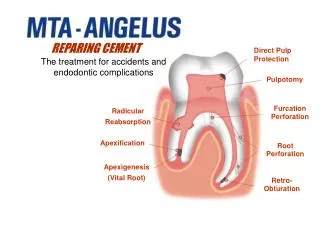
CLINICAL CONSIDERATIONS • The wide pulp chamber in the tooth of a young person will make a deep cavity preparation hazardous. • If opening a pulp chamber for treatment becomes necessary, its size and variation in shape must be taken into consideration. • The shape of the apical foramen and its location may play an important part in the treatment of root canals. • Pulpal-periodontal lesion due to accessory canals.
When filling materials contain harmful chemicals (e.g. acid in silicate cements and monomer in composites), an appropriate cavity liner should be used prior to the insertion of restorations. • The vitality of the pulp depends on its blood supply. The instruments called vitalometers test the reaction of the pulp to electrical or thermal stimuli.