Download
1 / 33
520 likes | 1.05k Views
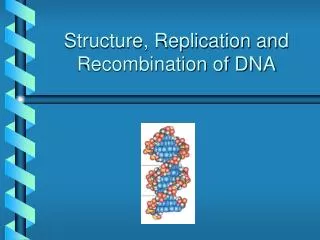
DNA Recombination. Roles Types Homologous recombination in E.coli Transposable elements. Biological Roles for Recombination. Generating new gene/allele combinations (crossing over during meiosis) Generating new genes (e.g., Immuno- globulin rearrangement)

E N D
DNA Recombination • Roles • Types • Homologous recombination in E.coli • Transposable elements
Biological Roles for Recombination • Generating new gene/allele combinations (crossing over during meiosis) • Generating new genes (e.g., Immuno- globulin rearrangement) • Integration of a specific DNA element • DNA repair
Practical Uses of Recombination 1. Used to map genes on chromosomes (recombination frequency proportional to distance between genes) 2. Making transgenic cells and organisms
Map of Chromosome I of Chlamydomonas reinhardtii cM = centiMorgan; unit of recombination frequency 1 cM = 1% recombination frequency Chlamydomonas Genetics Center
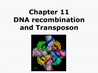
Types of Recombination • Homologous - occurs between sequences that are nearly identical (e.g., during meiosis) • Site-Specific- occurs between sequences with a limited stretch of similarity; involves specific sites • Transposition – DNA element moves from one site to another, usually little sequence similarity involved
Examples of Recombination Fig. 22.1
Holliday Model • R. Holliday (1964) • Holliday Junctions form during recombination • HJs can be resolved 2 ways, only one produces true recombinant molecules patch Fig. 22.2
EM of a Holliday Junction w/a few melted base pairs around junction Fig. 22.3
The recBCD Pathway of Homologous Recombination Part I: Nicking and Exchanging Fig. 22.5 a-e
recBCD Pathway of Homologous Recombination Part I: Nicking and Exchanging • A nick is created in one strand by recBCD at a Chi sequence (GCTGGTGG), found every 5000 bp. • Unwinding of DNA containing Chi sequence by recBCD allows binding of SSB and recA. • recA promotes strand invasion into homologous DNA, displacing one strand. • The displaced strand base-pairs with the single strand left behind on the other chromosome. • The displaced and now paired strand is nicked (by recBCD?) to complete strand exchange.
recBCD Pathway of Homologous Recombination Part II: Branch Migration and Resolution Fig. 22.5 f-h
recBCD Pathway of Homologous Rec. Part II: Branch Migration and Resolution • Nicks are sealed Holliday Junction • Branch migration (ruvA + ruvB) • Resolution of Holliday Junction (ruvC)
RecBCD : A complex enzyme • RecBCDhas: • Endonuclease subunits (recBCD) that cut one DNA strand close to Chi sequence. • DNA helicase activity (recBC subunit) and • DNA-dependent ATPase activity • unwinds DNA to generate SS regions Activity 2 and 3 linked
RecA • 38 kDa protein that polymerizes onto SS DNA 5’-3’ • Catalyzes strand exchange, also an ATPase • Also binds DS DNA, but not as strongly as SS Fig. 22.6
RecA Function Dissected • 3 steps of strand exchange: • Pre-synapsis: recA coats single stranded DNA (accelerated by SSB, get more relaxed structure, Fig. 22.8) • Synapsis: alignment of complementary sequences in SS and DS DNA (paranemic or side-by-side structure) • Post-synapsisorstrand-exchange: SS DNA replaces the same strand in the duplex to form a new DS DNA (requires ATP hydrolysis)
RuvA and RuvB • DNA helicase that catalyzes branch migration • RuvA tetramer binds to HJ (each DNA helix between subunits) • RuvB is a hexamer ring, has helicase & ATPase activity • 2 copies of ruvB bind at the HJ (to ruvA and 2 of the DNA helices) • Branch migration is in the direction of recA mediated strand-exchange
RuvC : resolvase • Endonuclease that cuts 2 strands of HJ • Binds to HJ as a dimer • Consensus sequence: (A/T)TT (G/C) - occurs frequently in E. coli genome - branch migration needed to reach consensus sequence!
RuvC bound to Holliday junction Fig. 22.31a
Transposable Elements (Transposons) • DNA elements capable of moving ("transposing") around the genome • Discovered by Barbara McClintock, largely from cytogenetic studies in maize, but since found in most organisms • She was studying "variegation" or sectoring in leaves and seeds • She liked to call them "controlling elements“ because they effected gene expression in myriad ways
Mutant Kernel Phenotypes • Pigmentation mutants • affect anthocyanin pathway • elements jump in/out of transcription factor genes (C or R) • sectoring phenotype - somatic mutations • whole kernel effected - germ line mutation • Starch synthesis mutants • - stain starch with iodine, see sectoring in endosperm
Some maize phenotypes caused by transposable elements excising in somatic tissues. Start with mutant kernels defective in starch synthesis (endosperm phenotypes) or anthocyanin synthesis (aleurone and pericarp phenotypes).
Somatic Excision of Ds from C Wild type Mutant Sectoring Fig. 23.19
Other Characteristics of McClintock's Elements • Unstable mutations that revert frequently but often partially, giving new phenotypes. • Some elements (e.g., Ds) correlated with chromosome breaks. • Elements often move during meiosis and mitosis. • Element movement accelerated by genome damage.
Molecular analysis of transposons • Transposons isolated by first cloning a gene that they invaded. A number have been cloned this way, via "Transposon trapping“. • Some common molecular features: • Exist as multiple copies in the genome • Insertion site of element does not have extensive homology to the transposon • Termini are an inverted repeat • Encode “transposases” that promote movement • A short, direct repeat of genomic DNA often flanks the transposon : “Footprint”
Ac and Ds • Ds is derived from Ac by internal deletions • Ds is not autonomous, requires Ac to move • Element termini are an imperfect IR • Ac encodes a protein that promotes movement - Transposase • Transposase excises element at IR, and also cuts the target
Structure of Ac and Ds deletion derivatives Ds is not autonomous, requires Ac to move! Fig. 23.20
How duplications in the target site probably occur. Duplication remains when element excises, thus the Footprint.
Mutator (A Retrotransposon) • Discovered in maize; differs significantly from Ac by structure and transposing mechanism • Autonomous and non-autonomous versions; many copies per cell • contains a long terminal IR (~200 bp) • transposes via a replicative mechanism, instead of a gain/loss mechanism • A “retrotransposon” • Similarities with retroviruses • move via an RNA intermediate • encode a reverse transcriptase activity
Control of Transposons • Autoregulation: Some transposases are transcriptional repressors of their own promoter(s) • e.g., TpnA of the Spm element • Transcriptional silencing: mechanism not well understood but important; correlates with methylation of the promoter
Biological Significance of Transposons • They provide a means for genomic change and variation, particularly in response to stress (McClintock’s "stress" hypothesis) (1983 Nobel lecture, Science 226:792) • or just "selfish DNA"? • No known examples of an element playing a normal role in development.