Download
1 / 84
860 likes | 1.21k Views
Atrial fibrillation and flutter: Practical Management Tips. Internal Medicine Residency Program Noon Conference 2011. Learning Goals. A brief discussion of supraventricular tachycardia (SVT) Review of AF and AFl physiology and EKG differentiation Management of atrial fibrillation (AF)

E N D
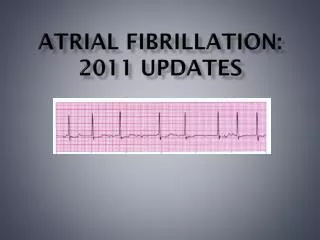
Atrial fibrillation and flutter:Practical Management Tips Internal Medicine Residency Program Noon Conference 2011
Learning Goals • A brief discussion of supraventricular tachycardia (SVT) • Review of AF and AFl physiology and EKG differentiation • Management of atrial fibrillation (AF) • Management of atrial flutter (AFl)
Management of fib/flutter • Anticoagulation • Cardioversion • Rate control • Rhythm control • Ablation • Cardiothoracic surgery
But, first, a brief diversion • Definition of supraventricular tachycardia (SVT) • Differentiating among types of SVT • Differentiating AF from AFl
Supraventricular tachycardia • Abbreviated SVT • “Supra” means “above” • Supraventricular tachycardia comes from above the ventricles • DO NOT CONFUSE with NSVT (non-sustained ventricular tachycardia) • (Essentially) all narrow-complex tachycardia has a supraventricular origin
SVT possible sites of origin • Sinus node • Atria • Atrioventricular node • His bundle • Or some combination of the above
Supraventricular tachycardia • Sinus tachycardia • Multifocal atrial tachcyardia • Paroxysmal atrial tachycardia • AV nodal reentrant tachycardia (AVNRT) • Atrioventricular reentrant tachycardia (AVRT) • Atrial fibrillation • Atrial flutter
Rare types of SVT • Inappropriate sinus tachycardia (previously called SNRT or SANRT) • Junctional ectopic tachycardia (JET) and paroxysmal junctional reciprocating tachycardia (PJRT) -- mostly seen in infants and children • Nonparoxysmal junctional tachycardia (NPJT) -- seen in acute right coronary artery occlusion and digitalis toxicity
Supraventricular tachycardia • A nonspecific term that technically refers to seven separate diagnoses • When clinicians use the term “SVT,” they mean that it is a narrow-complex tachycardia, and they cannot specify which one • Do not use the term “SVT” if you have an actual diagnosis • Do use “SVT” when the patient has “supraventricular tachycardia, not otherwise specified”
Supraventricular tachycardia • Sinus tachycardia • Multifocal atrial tachcyardia • Paroxysmal atrial tachycardia • AV nodal reentrant tachycardia (AVNRT) • AV reentrant tachycardia (AVRT) • Atrial fibrillation • Atrial flutter
When in doubt, use a vagal maneuver • Adenosine 6mg IV push -We don’t use enough adenosine! -But must be done with proper monitoring • Valsalva maneuver -Safe; usually ineffective • Carotid sinus massage -In properly selected populations, complications are rare
Carotid Sinus Massage Contraindications • Carotid bruit • Prior stroke or transient ischemic attack, unless imaging has shown no significant carotid disease • Myocardial infarction in the previous six months • History of serious cardiac arrhythmias (VT, VF)
Vagal maneuvers • Diagnostic -Usually you can learn which SVT it was by doing a vagal maneuver • Therapeutic -Vagal maneuvers can terminate AVRT and AVNRT
Differentiating AF from AFl • AFl is a macroreentrant atrial rhythm with a reentry circuit that involves a large area of atrial myocardium • AF is caused by multiple wandering wavelets, a hodgepodge of microreentrant circuits, often located in the pulmonary veins
Atrial flutter • P waves exhibit a “sawtooth” pattern referred to as flutter waves or “F” waves • Atrial rate is typically 250-350 beats per minute (bpm)
Atrial flutter, continued • Classically, atrial rate is 300 bpm with 2:1 AV conduction, leading to a heart rate of 150 bpm • But focus on the atrial pattern when diagnosing SVT--try to ignore the QRS complexes, just looking at the P (or F) waves at first
Atrial fibrillation • Rapid and irregular atrial activity at a rate of 350-600 impulses per minute • Usually irregularly irregular ventricular response • There are no P waves • Sometimes the F waves are so fine, the surface EKG cannot detect them
Atrial fibrillation terms • Paroxysmal • Persistent • Permanent • “Lone”
Atrial fibrillation terms, cont. • Paroxysmal -episodes terminate spontaneously in less than seven days • Persistent -fails to terminate within seven days
Atrial fibrillation terms, cont. • Permanent -AF lasts for more than one year, and -Cardioversion has not been attempted or has failed • “Lone” -patients less than 60 years of age without structural heart disease
Atrial fibrillation terms, cont. • This classification applies only when no clear reversible cause of AF. • If AF is clearly due to heart surgery, pericarditis, myocardial infarction, hyperthyroidism, pulmonary embolism, or other reversible causes, avoid this classification system
Management of fib/flutter • Anticoagulation • Cardioversion • Rate control • Rhythm control • Ablation • Cardiothoracic surgery
Anticoagulation (AC) • Recommendations are essentially the same for AF and AFl • First, assess if the patient is high risk for cardioembolic stroke • Most patients with high risk should be on AC if they ever were seen in AF or AFl
High risk for cardioembolic stroke • Rheumatic mitral stenosis -Mitral valve area less than 2.0 cm2 • Prosthetic heart valves • Hyperthyroid (?) -2006 ACC/AHA/ESC guidelines recommend INR 2-3 in all patients until euthyroid; ACCP does not comment on this
CHADS2 score • For use in patients without the high risk factors on the previous slide • There are other risk models, including the CHADS2-VASc score
When can you stop AC? • If AF was due to a completely reversed reversible cause, maybe you can stop • For AFl, you can never stop • After ablation you still can’t stop • If you had HF, and your EF returns to normal, you still can’t stop • Paroxysmal has same risk as others
When to stop AC • Pretty much you have to develop a contraindication to anticoagulation before you stop it • Fall risk is a relative contraindication; it’s rare that someone is falling so frequently that it rises to the level of contraindication
When to stop AC • Really, paroxysmal AF has same stroke risk as other AF • Caution when diagnosing AF! If you misdiagnosed NSR with PAC’s or multifocal atrial tachycardia as AF, you can doom someone to lifelong AC
When to bridge with heparin • If patient has any evidence of hypercoagulable state • If patient has prosthetic heart valves • Otherwise, risk of intracranial bleeding and HIT outweighs benefit of reduced stroke risk and warfarin skin necrosis
Risk of stroke per day off AC • Less than 4 events per 100 person-years prevented • One day is 4/100/365=0.0011% per day stroke risk reduction Efficacy and safety of anticoagulant treatment in acute cardioembolic stroke: a meta-analysis of randomized controlled trials. Paciaroni M, Agnelli G, Micheli S, Caso VS. Stroke. 2007;38(2):423.
Take-home point • Warfarin>ASA+clopidogrel>ASA • This applies to stroke prevention • And to bleeding risk • Every AF patient who is off warfarin should be on aspirin unless there is a contraindication to ASA therapy
Clopidogrel + Warfarin = Bleed • ASA+warfarin and ASA+clopidogrel are relatively safe in most situations where both are indicated • Warfarin+clopidogrel has a relative risk of bleeding >3 times greater than warfarin alone • ASA+warfarin+clopidogrel has less rigorous data; evidence suggests >5 times greater than asa+clopidogrel alone
Dabigatran and Apixaban • Oral anticoagulant medications that do not require monitoring • Dabigatran approved for AC in AF/AFl in USA; NYS Medicaid and NBHN do not pay for it. Medicare part D does pay • Apixaban likely similar; not yet approved
You’re the night intern • 78 W with HTN, DM, COPD, and history of paroxysmal AF • Nurse pages you to say that HR is now 172 beats per minute after albuterol • You ask, “what’s the blood pressure?” • The nurse says she will check. You go to the bedside.
You’re the night intern • HR=172 irregular, BP is 72/48 • Pt. is pale, diaphoretic, lethargic but responds to questions • You confirm with EKG that the rhythm is atrial fibrillation • Put patient in Trendelenberg position to optimize cerebral perfusion, then you…
Single best answer: Next step • A) DC cardioversion without sedation • B) Ativan 2mg IV over 30 seconds, then direct current (DC) cardioversion • C) Amiodarone 150mg IV over 15 min. • D) Diltiazem 10mg IV over 2 minutes • E) Adenosine 6mg IV push