Download
1 / 1
10 likes | 142 Views
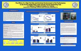
The Effect of a High-Fat Meal On Microvascular Function. V Lee, B Martin, M Ikanovic , TJ Anderson Department of Cardiac Sciences, Libin Cardiovascular Institute of Alberta, University of Calgary, Calgary, AB, Canada. Introduction. Results.

E N D
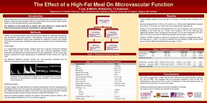
The Effect of a High-Fat Meal On Microvascular Function V Lee, B Martin, M Ikanovic, TJ Anderson Department of Cardiac Sciences, Libin Cardiovascular Institute of Alberta, University of Calgary, Calgary, AB, Canada Introduction Results High-fat diets are known to be associated with the development of atherosclerosis over a lifespan. A high-fat meal has been shown to acutely impair macrovascular health, but the impact on microvascular function is less well studied. The objective of this study was to assess the acute effects of a single high-fat meal on microvascular function, a barometer of vascular health. Twenty healthy subjects (mean age 22.9 ± 4.8 years, 8 females) were included in this study. Glucose decreased from 4.80 to 4.40 mmol/L (p = 0.003) while triglycerides increased from 0.84 to 1.41 mmol/L (p < 0.001) two hours following the high-fat meal. Analysis of VTI showed that it decreased from 147 ± 38 cm fasting to 122 ± 28 cm post-prandially on the meal day (p = 0.010). This change in VTI on the meal day was significantly different when compared to the change in VTI on the control day (VTI 134 ± 36 to 134 ± 28 cm) (p-value for differences between the two days = 0.040). Analysis of FMD revealed no significant changes from fasting to postprandial state on the meal day (p = 0.891). This was no different from the change in FMD on the control day (p-value for differences between the two days = 0.273) Group A (n=10) Group B (n=10) Methods ▪ ▪ ▪ ▪ ▪ Healthy, non-smoking subjects were recruited and screened for cardiovascular disease, high cholesterol, high blood pressure, and diabetes. Using brachial artery ultrasound, microvascular endothelial function was assessed by hyperemic velocity time integral (VTI) while macrovascular endothelial function was assessed by flow-mediated dilation (FMD). Study Design In a randomized crossover design, subjects were fed a high-fat commercial breakfast meal (860 calories, 50g of fat) on the meal day, receiving endothelial function assessment pre-prandial and reassessment two hours postprandial. On the control day, subjects received the same assessments of endothelial function without the high-fat meal. The difference between vascular function pre- and post-meal compared with the difference in measures on the control day was assessed. Statistical Analyses For each subject, the delta between the baseline assessment and the reassessment of endothelial function two hours later was calculated on both the control day and the meal day. Differences between the deltas on the control day and meal day were then analyzed using paired student t testing. Analysis was done using SPSS Statistics 17.0. Using the crossover design including 20 subjects, we had 80% power to detect a 25% clinically significant difference in VTI, our primary endpoint. ▪ ▪ ▪ ▪ ▪ Table 2: Comparisons of Endothelial Function Figure 1: Randomized Crossover Design Table 1: Characteristics of Study Population (a) (b) Conclusions (a) Measurement of velocity time integral for the first Doppler wave in reactive hyperemia. (b) Measurement of brachial artery diameter at rest and during reactive hyperemia. Our results suggest that a single high-fat meal affects the microvascular bed, impairing VTI, but not FMD. This suggests that the association between a high fat diet and atherosclerosis may also be due in part to impairment of the microvascular bed, an impact that can be seen in an acute time frame. Funding provided by the Heart and Stroke Foundation of Canada. The authors have no disclosures relevant to this poster.