Download
1 / 67
670 likes | 809 Views
Central nervous system malignancies. Commonest malignant solid tumors in childhood 20% of cancers in age < 15 years Annually 20 – 26/ 1 million children below the age of 16 years

E N D
Commonest malignant solid tumors in childhood 20% of cancers in age < 15 years Annually 20 – 26/ 1 million children below the age of 16 years Age –stratified incidence is: <1year - 27/ 1 million 1 – 4 - 31/ 1 million 5 – 9 - 27/ 1 million 10 – 14 - 20/ 1 million Slightly higher frequency in boys 1.25:1 (especially for medulloblastoma and germinoma) Epidemiology
Etiology and pathogenesis Association between primary CNS tumors and following conditions/ genetic disorders : 1. Neurofibromatosis (NF) type 1 and 2 2. Tuberous sclerosis 3. Von Hippel- Lindau syndrome 4. Gordlin’s, Cowden’s, Turcot’s syndromes 5. Li-Fraumeni syndrome (mutation of suppressor oncogene p53) Deletion of chromosome 17 or 20 (medulloblastoma) Exposition of the brain to ionizing radiation i.e. after cranial radiotherapy in leukemia
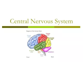
Pathology Supratentorial lesions (30 – 40%): 1. Cerebral hemisphere ( astrocytoma, ependymoma, glioblastoma, meningioma) 2. Sella or chiasm ( craniopharyngioma, pituitary adenoma, optic nerve glioma) Infratentorial lesions (60 – 70%) 1. Cerebellum (medulloblastoma, astrocytoma, meningioma) 2. Brain stem ( astrocytoma, ependymoma, glioblastoma)
Classification Based on histogenesis and predominance of cell type Degree of malignancy is defined by grading system e.i. WHO grade based on cellular morphology, mitotic index, anaplasia and necrosis: grades I and II represent benign tumors grades III and IV - malignant tumors
Clinical presentation Depends on : -age -anatomical site -tumor type -raised intracranial pressure (ICP) -localizing neurological deficits
Signs of increased ICP Direct tumor infiltration Compression of normal structures Secondary to obstruction of the cerebrospinal fluid (CSF)
Older children: -inilially behavioural changes and declining school performance prior to development of the more classical features of headache, nausea and vomiting , headaches start as generalized and intermittent > increase in both intensity and frequency with time - the child may awake with headache at night, with the pain generally being worse in the morning and improving during the day with an upright posture -School-age children complain of visual disturbances
Infants and younger children: plasticity of the developing skull and inability to communicate symptoms > -infant may be irritable, with failure to thrive, associated with anorexia and vomiting -regression of developmental milestones -increase head circumference with widened sutures and a tense anterior fontanelle „sun-setting” sign
Symptoms and signs according to anatomical site of CNS tumors Supratentorial (30-40%) Cerebral hemisphere- hemiparesis, spasticity, seizures (focal or generalized) - para/suprasellar – endocrinopathy (growth failure, diabetes insipidus, pubertal abnormality) - hypothalamus – diencephalic syndrome (infants), developmental and behavioural abnormalities - optic pathway – visual field acuity, color vision deficits, optic atrophy, nystagmus, head tilt - pineal - Parinaud’s syndrome, sleep abnormalities - thalamus, basal ganglia – pain, sensory loss, memory disturbances - intraventicular - meningeal
Infratentorial (60 – 70%) - posterior fossa – ataxia, nystagmus, dysmetria (presents as clumsiness or worse handwriting) - brainstem – multiple cranial nerve palsies, hemiparesis, spasticity, mood changes Spinal (2 – 5%) - primary intramedullary – pain (local back and root pain), motor and sensory disturbance - spinal metastases – scoliosis, sphincter (bowel, bladder) disturbances, reflex changes
Diagnostic evaluation Magnetic resonance and computed tomography – basic imaging techniques for brain tumors Positron emission tomography – help to distinguish tumors or lesions with a volume greater than 1 cm3 Conventional radiography of the skull: bone structure, separating off sutures ( due to ICP), calcification within the brain Special methods (for special indications): - brain scintigraphy - angiography -ultrasonography -myelography
Additional diagnosis Cerebral fluid analysis ( to determine spread of the tumor to the spinal fluid) Electroencephalography Stereotactic biopsy
Therapy Neurosurgery for maximum tumor removal and low morbidity depending on the location and extent of the tumor -often preoperative relief of intracranial pressure by ventriculoperitoneal or ventriculoarterial shunt - preoperative reduction of tumor edema by corticosteroids - in patients with seizures - anticonvulsive therapy
Radiotherapy – extension and volume of irradiation depend on the biology and histology of the tumor, age of the child and combination with chemotherapy and neurosurgery - irradiation in children < 3 years of life only in special cases Chemotherapy – depends on tumor type, location and age -efficacy and penetration depend on vascularization of the tumor
Malignant, embryonal tumor derived from precursor cells of sympathetic ganglia and adrenal medulla Other types of tumors derived from sympathetic nervous system: -ganglioneuroblastoma -ganglioneuroma -pheochromocytoma Possibility to spontaneous regression and differentiation to benign tumor in infants less 1 year of age extremely malignant in older children
Epidemiology 8% of all neoplasms in children Most frequent malignant neoplasm in infants Mean age at diagnosis 2 – 5 years
Pathology Two distinct entities: Infant: 1. possibility of spontaneous regression (apoptosis or differentiation into ganglioneuroblastoma) 2. chemosensitive, chemocurable Older3. chemoresistant malignancies
Molecular cytogenetics MYCN oncogene amplification. MYCN is located on chromosome 2p. Independent prognostic factor: in stage III EFS for patients with a single copy is curable 80%; for those with amplification MYCN – 20% DNA ploidy: hyperploidy = good prognosis Nerve growth factor receptor: ligands for high –affinity tyrosine kinase receptors TRKA, TRKB, TRKC: -TRKA expression is associated with MYCN single copy, low stage and good prognosis - TRKA (-) + MYCN amplification= very poor survival Structural and numerical abnormalities of chromosome 1
Clinical manifestation Occurence in any area with sympathetic nervous system Primary location: -abdomen 65% -adrenal medulla or sympathetic ganglia 46% -posterior mediastinum 15% -pelvic 4% -head and neck 3% -others 8%
Common symptoms Weight loss Fever Abdominal disturbances Irratability Pain of bones and joints Child not stand up, not walk Pallor Lassitude
Symptoms associated with catecholamine production Paroxysmal attacks of sweating, flushing, pallor Headache Hypertension Palpitation
Paraneoplastic syndromes VIP syndrome: untreatable diarrhea,with low level of potassium Opsoclonus- myoclonus Anemia, trombocytopenia, leukopenia ( in bone marrow infiltration or massive hemorrhage)
Local symptoms Abdomen: -intra-abdominal tumor –paravertebral and presacral -neurological dysfunction -abdominal distension Liver: -hepatomegaly Chest, posterior mediastinum, vertebrae: -compression of trachea > coughing, dyspnea -infiltration in vertebral foramina > dumbbell tumor -compression of nerves >disturbances of gait, muscle weakness, parasthesia, bladder dysfunction, constipation
Eyes: -periorbitaledema, swelling, yellow- brownecchymoses -proptosis and exophthalmos, strabismus, opsoclonus -papillaryedema, bleeding of the retina, atrophy of the opticnerve Neck: -cervicallymphadenopathy -supraclavicular tumor -Hornersyndrome: enophthalmos, miosis, ptosis, Racconeyes
Skin: -subcutaneous nodules of blue color > reddish > white owing to vasoconstriction from release of catecholamines after palpation - nodules are mainly observed in neonates or infants with disseminated NBL Bone: -pain involvement mainly in the skull and long bones - in X-rays – lytic defects with irregular margins and periosteal reaction Bone marrow: -trombocytopenia, anemia
Metastases Lymphatic and/or hematogenous spread Often initially present in children (40 – 50% children < 1 year and 70% children > 1 year) Metastatic spread mostly in bone marrow, bone, liver, skin
International Staging System for NBL(INSS) 1 - localized tumor with complete excision,lymph nodes negative 2a - localized tumor without incomplete gross excision, representative, ipsilateral nonadherent lymph nodes negative for tumor microscopically 2b – ipsilateral nonadherent lymph nodes positive for tumor. Enlarged contralateral lymph nodes negative microscopically 3 – unresectable unilateral tumor infiltrating across the midline,with or without regional lymph node involvement or localized unilateral tumor with contralateral regional lymph node involvement or – midline tumor with bilateral extension by infiltration or by lymph node involvement 4 – any primary tumor with dissemination to distant lymph nodes, bone, bone marrow, liver, skin or other organs (except as defined for stage 4S 4s localized primary tumor (as defined for stages 1, 2a, 2b) with dissemination limited to skin, liver or bone marrow; limited to infants aged less than 1 year)
Laboratory findings Tumor markers: -catecholamines: vanillylmandelic acid (VMA), homovanillic acid (HVA) dopamine in urine/ plasma adrenaline, noradrenaline -neuron-specific enolase (glycolitic enzyme of brain and neuroendocrine tissues) -NSE Ferritin Lactate dehydrogenease (LDH) Bone marrow (aspiration and biopsy)
Locoregional involvement Computed tomography scan and/or Ultrasound and/or magnetic resonance imaging → localize the mass, provide measurements, give anatomical information about intra- and extraperitoneal structures, differentiate cystic from solid tumors, define the extent of a primary tumor and its relationship with other structures, detect small calcification
Evaluation of metastases Bone marrow metastases – bone marrow aspiration and trephine biopsy Skeletal metastases - X-ray, Tc-99 scintigraphy, mIBG scintigraphy demonstrates primary, residual tumor masses,diffuse bone marrow infiltration, skeletal, lymph node and soft tissue metastases FDG-PET scanning
Therapy Depends on: age, stage, localization and molecular features at diagnosis Surgery Chemotherapy IV stage Radiotherapy Target radiotherapy (I-131-mIBG) Differentiation therapy (retinoids 13-cis and all-trans) Immunotherapy: anti-GD2 antibodies Auto-BMT
Prognosis Depends on: -age (favorable if less than 18 months of age at diagnosis), -stage and localization (favorable in primary NBL of thorax, presacral and cervical) -involvement of lymph nodes (poor prognosis) Low-risk group - 90% long-term survival Intermediate and high-risk groups: -response to initial treatment 60-70% of children with complete or partial remission -after consolidation therapy (high-dose chemotherapy+ autologous stem cell support) –EFS after 3 years is 40-60%
Malignant: -hepatoblastoma 43% -hepatocellular carcinoma 23% -sarcoma 6% Benign: -vascular (haemangioma,haemangioendothelioma) 13% -hamartoma 6% -others 9%
Incidence (malignant) -1.2 –5% of all neoplasms in childhood Boys: girls 1.4 –2 :1 High incidence in genetically associated syndromes: -Beckwith-Wiedemann syndrome, familial adematous polyposis, trisomy 18, glycogen storage disease, hereditary tyrosinemia, Li-Fraumeni syndrome HBL –mostly in infants, rarely after the age of 3 years Hepatocellular Ca – in older children, in adolescents, after hepatitis B
Clinical symptoms Abdominal mass and/ or abdominal distention Anorexia, weight loss Lethargy Jaundice and ascites Abdominal pain Pallor Precocious puberty (hepatoblastoma), virilization (due to gonadotrophin production by the tumor) Generalized osteoporosis (HBL)- tumor cells secrete osteoclast-activating factor
Laboratory diagnosis Serum AFP elevated in 80 - 90% of children with HBL and in 60-70% with hepatocellular Ca B-HCG elevated in both Increased bilirubin Increased serum aspartate aminotransferase (AST), alanine transaminase (ALT) because of associated hepatitis or cirrhosis Anemia, thrombocytosis, trombocytopenia (rarely) biopsy
Radiological diagnosis Ultrasound – liver enlarged, displacement of stomach and colon, elevated diaphragm on the right side CT, MRI Liver scintigraphy metastases: chest X-ray, lung CT HBL hepatocellular carcinoma
Treatment Presurgical chemotherapy (makes HBL resectable) Complete resection (initially > 50% of liver tumors are not totally resectable) Liver transplantation Prognosis: Depends on stage of the tumor after complete tumor resection and chemotherapy 65-75 % children with HBL and 40-60% with hepatocellular Ca survive
Develop from embryonal germ cells Represent ectodermal, endodermal and mesodermal lineages Approximately 3% of childhood malignancies Annually 2.4 – 3.8/ 1 million/per year 2/3 GCTs in children occur in extragonadal sites The commonest GCT - sacrococcygeal teratoma Bimodal age distribution: one peak in children < 3y ears of age (sacrococcygeal tumors, yolk sac tumors of testis) - the later- the later GCT of the ovary, testis, and intracranial sites
Histological classification of gonadal and extragonadal tumors Germ cell and germinoma/ dysgerminoma and embryonal yolk sac tumor (pluripotent cells) a)extraembryonic structures - yolk sac or endodermal sinus tumor - choriocarcinoma b) embryonal ecto-, meso-, endodermal origin tissues represented -teratoma c) embryonal carcinoma Gonadal germ cells and stroma tumor (Sertoli and Leydig cells) Epithelial cells (ovarian origin) and granulosa cell tumor or mixted form as well as epithelial cell tumors more common in adults