Download
1 / 1
10 likes | 142 Views
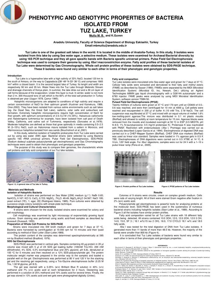
PHENOTYPIC AND GENOTYPIC PROPERTIES OF BACTERIA ISOLATED FROM TUZ LAKE, TURKEY Mutlu M. B. and K.Guven Anadolu University, Faculty of Science Department of Biology Eskisehir, Turkey Email:mbmutlu@anadolu.edu.tr

E N D
PHENOTYPIC AND GENOTYPIC PROPERTIES OF BACTERIA ISOLATED FROM TUZ LAKE, TURKEY Mutlu M. B. and K.Guven Anadolu University, Faculty of Science Department of Biology Eskisehir, Turkey Email:mbmutlu@anadolu.edu.tr Tuz Lake is one of the greatest salt lakes in the world. It is located in the middle of Anatolia-Turkey. In this study, 9 isolates were isolated from this lake by using Sea water agar, a selective medium. These isolates were examined for Archaeal/Bacterial diversity by using 16S PCR technique and they all gave spesific bands with Bacteria spesific universal primers. Pulse Field Gel Electrophoresis technique was used to compare their genome by using Xba I macrorestriction enzyme. Fatty acid profiles of these bacterial isolates of Tuz Lake were determined by Gas Chromotography. Whole cell protein profiles of these isolates were obtained by SDS-PAGE technique. These 9 isolates were found very similar to each other in terms of their phenotypic and genotypic properties. Introduction Tuz Lake is a hypersaline lake with a high salinity of 33% NaCl, located120 km to the south of Ankara, on the way to Cappadocia (38o 48o N-33o 36o E) and comprises 1665 km2 within a closed basin. It is the second largest lake of Turkey. Its lenght and width are respectively 90 km and 35 km. Water flows into the Tuz Lake through Melendiz Stream and drainage channels of Konya plain. In summer, the lake dries out and a 30 cm layer of salt forms because of the evaporation. Under this layer is mud. In winter, water is not more than 2 m. 300 thousand tons of salt which is the 60% of the total salt production in Turkey is obtained per year (Uygun 1978, Çamur 1995). Halophilic microorganisms are adapted to conditions of high salinity and require a certain concentration of NaCl for their optimum growth (Kushner and Kamekura, 1988; Oren,2002). They have been isolated from various saline environments such as salt lakes (eg. the Dead Sea, the Great Salt Lake), salterns, solar salts and subsurface salt formation. Extremely halophilic microorganisms require high concentration of NaCl for their growth, with optimum concentrations of 2.5–5.2 M (15–30%). Haloarcula vallismortis and Haloterrigena turkmenica for example, have been isolated from salt pool of Death Valley, California, and saline soil of Turkmenia, respectively (Gonzales et al.,1978). Moderate halophiles are defined as those that grow optimally in media containing 0.5–2.5 M (3–15%) NaCl, such as Halomonas maura isolated from a saltern in Morocco, and Marinococcus halophilus isolated from sea sands (Bouchotroch et al.,2001) In this study, selective isolation of halophilic prokaryotes from Tuz Lake were carried out by using sea water (SW) agar. Bacterial strains were selected by using Bacteria specific 16S rRNA universal primers. Pulse Field Gel Electrophoresis, SDS-PAGE (Sodium dodecyl sulphate polyacrylamide gel electrophoresis) and Gas Chromotography techniques were used to obtain their phenotypic and genotypic properties. The purpose of this study was to compare their genomes, the characteristic whole cell proteins and fatty acid composition of our Tuz lake isolates. Fatty acid composition Tuz Lake isolates were inoculated onto Sea water agar and grown for 7 days at 37 oC. Cellular fatty acids were extracted and derivatized to their fatty acid methyl esters (FAME) as described by Sasser (1990). FAMEs were separated by the MIDI (Microbial Identification System) (Microbial ID, Inc, Newark, Del.) utilizing an Agilent Technologies 6890N gas liquid chromatography with a G2614A autosampler and a 6783 injector. FAME peaks were analysed by using MIDI Microbial Identification System, software version TSBA 50. Pulsed Field Gel Electrophoresis (PFGE) Twenty mililiters of cultures were grown at 37 oC and 170 rpm until an OD600 of 0.5–0.6 was reached, and were then centrifuged for 10 min at 4000 g. Cell pellets were washed and resuspended in 200 µl of buffer A (10 mM Tris, 3 M NaCl). The cell suspension was equilibrated at 37 oC and mixed with an equal volume of melted 1.6% low-melting-point agarose.The mixture was distributed in 0.1 ml plastic moulds (BioRad) and allowed to solidify at room temperature for 15 min. Agarose blocks were removed from the moulds and incubated overnight at 50 oC in ESP (0.5 MEDTA, pH 9–9.5; 1% N-laurylsarcosine; 0.5 mg/ml proteinase K). Proteinase K was inactivated with PMSF. Restriction with endonucleases (Xba I, Promega) was carried out as previously described (Lopez-Garcia et al. 1994). Electrophoresis of digested DNA was carried out in a CHEF-Mapper System (BioRad). CHEF DNA size markers (BioRad) was used as linear size standard. Samples were loaded in 1% agarose gels in 0.5·X TBE. Electrophoreses of DNA digestions were carried out in 0.5 XTBE at 14 oC, 6 V/cm, 120o field angle. For Xba I digestions, samples were run for 24 h with a 1–10 s pulse linear ramp (Pena et al., 2005). Figure 1-2. A general view of Tuz lake in Turkey. Figure 3. Protein profiles of Tuz Lake isolates Figure 4. PFGE patterns of Tuz Lake isolates Materials and Methods Isolation of Halophilic Bacteria Isolation of strains was performed on Sea Water [(SW) medium (g l-1): NaBr 0.65; NaHCO3 0.167; KCl 5; CaCl2 0.723; MgSO4 .7H2O: 49.49; MgCl2.6H2O 34.567; NaCl 195; yeast extract (YE), 1; agar, 20] (Rodriguez-Valera, 1985). Pure cultures were obtained by succesive single colony isolations with streak plate technique. Morphological and Cultural Characteristics 9 strains were choosen for the study. Isolated strains were examined for colony and cell morphology. Cell morphology was examined by light microscopy of exponentially growing liquid cultures. Gram staining was performed using acetic acid-fixed samples as described by Dussault (Dussault, 1955). Preparation of Whole Cell Proteins for SDS-PAGE Strains were inoculated into SW broth medium and grown for 7 days at 37 oC. Bacteria were harvested by centrifugation at 10.000 rpm for 10 minutes and then lysed according to method of Stan-Lotter (Stan-Lotter,1989). The protein content of the samples was determined spectrophotometrically at 595 nm by Bradford method (Bradford, 1976). SDS Gel Electrophoresis SDS-PAGE was performed in vertical gels. Samples containing 40 μg protein in 20 μl volume was mixed with 5 μl of 2X SDS gel loading buffer (100mM Tris-HCl, 200 mM dithiothreitol, 4% SDS, 0.2% bromophenol blue and 20% glycerol, pH 6.8), denatured at 100 oC for 5 minutes and then resolved on a 12% SDS-polyacrylamide gel. The protein molecular weight marker was prepared in the similar way to the samples and loaded a parallel well on the gel. Electrophoresis was performed at 80 V and 120 V for the stacking gel and the resolving gel respectively, using a Tris-Glycine electrophoresis buffer (0.25 M Tris, 1.92 M glycine, pH 8.5). The gel was stained with a 0.25% (w/v) Brilliant Blue R solution in 40% (v/v) methanol and 7% (v/v) acetic acid at room temperature for 2 hours. Destaining was performed in a solution of 25% methanol and 10% acetic acid for several times. Finally, the gel was stored in 13% acetic acid and wet gels were photographed digitally (Uvitec). Results Colonies of 9 strains were circular, cream on complex growth medium. Cells were rods of varying lenght. All of them were stained Gram negative after fixation in 2% (w/v) acetic acid. Polyacrylamide gel electrophoresis is powerful tools for analysing proteins at the molecular level. SDS-PAGE has been used in the systematics of numeous bacterial strains including halophilic isolates (Stan-Lotter et al., 1989). According to Figure 3 all the isolates have similar protein profiles. Fatty acid composition varied for all Tuz Lakestrains with 18 different fatty acids being detected. All strains contained 10:0 3OH, 12:0, 12:0 2OH, 12:0 3 OH, 14:0, 15:0, SF 3 ( 16:1 w7c/15 iso 2 OH), 16:0, 17:0 CYCLO 18:1 w7c and 18:0 fatty acids. Xba I was tested for the total digestion of DNA from Tuz Lake isolates. It generated more than 11 bands of more than 48.5 kb. However, the majority of the bands were smaller than 800 kb (Figure 4). It is concluded that Tuz Lake isolates of bacteria were found very similar to each other in terms of their phenotypic and genotypic properties. REFERENCES Bouchotroch S, Quesada E, del Moral A, Llamas I, Bejar V: Halomonas maura sp. nov., a novel moderately halophilic, exopolysaccharide-producing bacterium. Int J Syst Evol Microbiol 2001, 51:1625-32. Bradford, M. M. 1976. A rapid and sensitive method for the quantitation of microgram quantities of protein utilizing the principle of protein-dye binding. Analyt. Biochem. 72:248-254. Çamur, M. Z. and Mutlu, H. (1995). Geological Bulletin of Turkey. 38:67-73 Dussault, H. P. (1955). An improved technique for staining red halophilic bacteria. J Bacteriol 70, 484-485. Gonzalez C, Gutierrez C, Ramirez C: Halobacterium vallismortis sp. nov. an amylolytic and carbohydrate-metabolizing, extremely halophilic bacterium. Can J Microbiol 1978, 24:710-715. Kushner DJ, Kamekura M: Physiology of halophilic eubacteria. In Halophilic bacteria. Volume I. Boca Raton, CRC Press; 1988:109-140. Lopez-Garcia P, Anton J, Abad JP, Amils R (1994) Halobacterial megaplasmids are negatively supercoiled. Mol Microbiol 11:421–427 Oren A: Halophilic Microorganisms and their environments. Dordrecht, Kluwer Academic; 2002. Peña, A., Valens, M., Santos, F., Buczolits, S., Antón, J., Kämpfer, P., Busse, H.-J., Amann, R., Rosselló-Mora, R. 2005. Intraspecific comparative analysis of the species Salinibacter ruber. Extremophiles 9:151-161 Rodriguez-Valera, F., A. Ventosa, G. Juez, And J. Imhoff. 1985. Variation of environmental features and microbial populations in salt concentrations in a multi-pond saltern. Microb. Ecol. 11:107:115. Sasser, M. (1990): Identification of bacteria through fatty acid analyses. In Klement, Z., K., Rudolph, D. C. Sands (eds) Methods in phytobacteriology (pp 199-204) Academiai Kiado, Budapest, Hungary. Stan-Lotter, H., Lang, F. J. And Hochstein, L. I. 1989. Electrophoresis and isoelectric focusing of whole cell and membrane proteins from the extremely halophilic archaebacteria. Appl. And Theor. Electrophoresis 1:147-153. Uygun, A. and Şen, E. (1978). Geological Bulletin of Turkey. 21:113-120.