Download
1 / 18
220 likes | 426 Views
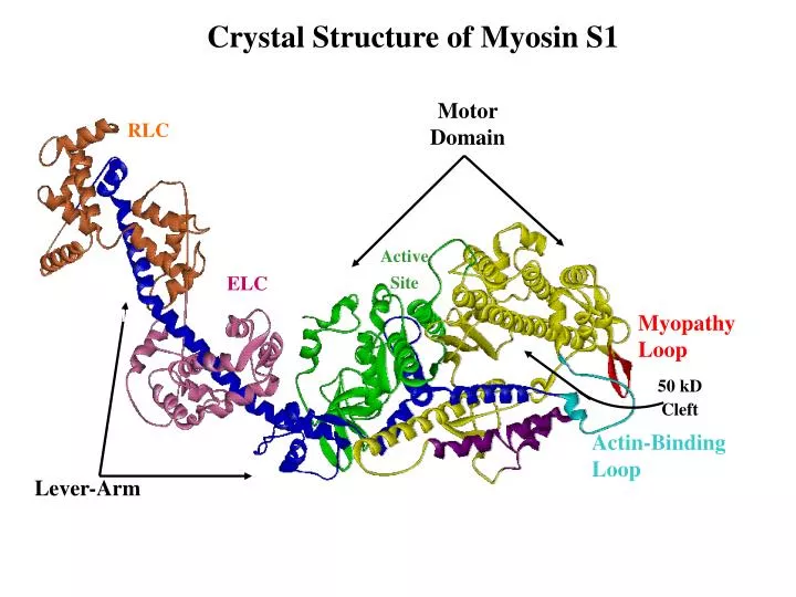
Crystal Structure of Myosin S1. Motor Domain. RLC. Active Site. ELC. Lever Arm. Myopathy Loop. 50 kD Cleft. Actin-Binding Loop. Helix-Loop-Helix. Lever-Arm. Pi, ADP. ATP. Hydrolysis. Structure/Function Relationships in Myosin. Hydrolyze ATP Bind Actin Generate Powerstroke

E N D
Crystal Structure of Myosin S1 Motor Domain RLC Active Site ELC Lever Arm Myopathy Loop 50 kD Cleft Actin-Binding Loop Helix-Loop-Helix Lever-Arm
Pi, ADP ATP Hydrolysis Structure/Function Relationships in Myosin • Hydrolyze ATP • Bind Actin • Generate Powerstroke • Coordinate Functions
Myosin: “A Whale of a Protein” Blow Hole Tail Mouth
Structure/Function Relationships in Myosin Lever-arm - generates powerstroke. Active Site - hydrolyzes ATP. Rigid Relay Loop - along with converter domain coordinates active site and lever arm. Actin-binding Cleft - mediates affinity for actin.
How Can the Details of Changes Be Established? Crystallograhy can provide some information about changes since myosin can be crystallized in different nucleotide states. However, to address DYNAMIC changes in the protein other methods are required which are 1) compatible with biological reaction conditions, 2) sensitive to structural changes, and 3) have excellent temporal resolution. Fluorescence Spectroscopy
2936 441 512 Skel. YLRKSPFDAKSSVFVVHPKES / EKM.FLWMVIRIN / KKEGIEWEFID Card. EAQTRPFDLKKDVFVPDDKQE / ERM.FNWMVTRIN / KKEGIEWTFID Dcty KLTVSDKRYIWYNPDPKERDS / GRL.FLWLVKKIN / LKEKINWTFID Smooth LAQA.DWSAKKLVWVPSEKHG / ERL.FRWILTRVN / QREGIEWNFID FF F F 546 597 625 Skel. / ILEEECMFPKATD / DYNISGWLEKNK / KTLALLFATY... Card. / ILEEECMFPKATD / DYNIIGWLQKNK / KLLSTLFANY... Dicty / LLDEQSVFPNATD / MYEIQDWLEKNK / NVVTKLFND.... Smooth / LLDEECWFPKATD / TYNASAWLTKNM / KFVADLWKDVDRI M F W
Upper 50 kDa Subdomain F425W ELC Actin-Binding Cleft V413W W546M Lower 50 kDa Subdomain
FHC – Familial Hypertrophic Cardiomyopathy R413Q mutation Normal
Steady-State Fluorescence Properties MAX(nm) 333 0.36 344 0.20 351 0.14
Acrylamide Quenching F0/F = 1 + KSV[Q] KSV (M-1) kq (M-1·ns-1) 11.5 6.4 5.3 1.7
Relative Fluorescence of 625 MDE Bound to Actin
Actin-Induced Conformational Changes Trp625 - Unchanged Trp546 -adopts a more buried conformation MAX (nm) MAX (nm) 334 344 333
Actin Bound Fluorescence: ADP-Bound vs. Rigor MAX 347 341 338 MAX 336 337
Steady-State Fluorescence of F425W-MDE Nucleotide Bound Peak Intensity MAX KSV (M-1·ns-1) None 100% 338 4.1 ± 0.02 MgADP 97% 339 4.1 ± 0.02 MgATP 80% 345 5.7 ± 0.06
Structural Model of Acto-Myosin Interactions A:M-ATP A:M-ADP A:M Actin V413W W546 Cleft Conformational Changes F425W Weak Binding Cleft Closure Motor Domain Rotation