Download
1 / 19
260 likes | 808 Views
Ischemic Heart Disease. Ms . Leonardo Roever. Heart - Pathology. Ischemic Heart Disease Hypoxemia (diminished transport of oxygen by the blood) less deleterious than ischemia Also called coronary artery disease (CAD) or coronary heart disease IHD =Syndromes

E N D
Ischemic Heart Disease Ms. Leonardo Roever
Heart - Pathology Ischemic Heart Disease • Hypoxemia (diminished transport of oxygen by the blood) less deleterious than ischemia • Also called coronary artery disease (CAD) or coronary heart disease • IHD =Syndromes • late manifestations of coronary atherosclerosis • Cause => 90% of cases, coronary atherosclerotic arterial obstruction
Heart Ischemic Heart Disease • Classification = mainly 4 types • Myocardial infarction (MI) • Sudden cardiac death • Angina pectoris • Chronic IHD with heart failure • Acute Coronary syndromes • important predisposing factor -Plaque disruption or Acute plaque change • Acute myocardial infarction • Unstable angina • Sudden cardiac death
Heart Ischemic Heart Disease • 75% stenosis = symptomatic ischemia induced by exercise • 90% stenosis = symptomatic even at rest • Pathogenesis • ↓ coronary perfusion relative to myocardial demand • Role of Acute Plaque Change • (Erosion/ulceration, Hemorrhage into the atheroma, Rupture/fissuring, Thrombosis) • Role of Inflammation • T cell, Macrophages (MMPs), CRP • Role of Coronary Thrombus • The most dreaded complication • Role of Vasoconstriction (VC) • Platelet & Endothelial factors, VC substances
Heart - Pathology Ischemic Heart Disease • Angina Pectoris • Chest discomfort = prolonged, recurrent, different qualities • Cause = transient myocardial ischemia( seconds to minutes) • Patterns • Stable = 75% vessel block, transient ( <15 minutes), aggravated by exertion, relived by rest & Nitroglycerin (VD) • Prinzmetal = coronary spasm, episodic, Typical EKG change – ST elevation, Relived by VD but not rest • Unstable = 90% vessel block or Acute plaque change ( superimposed thrombus), prolonged ( >15 min.), not relived by rest, VD, Pre-infarction Angina
Transmural Full thickness Superimposed thrombus in atherosclerosis Focal damage Sub-endocardial Inner 1/3 to half of ventricular wall Decreased circulating blood volume( shock, Hypotension, Lysed thrombus) Circumferential MI - Types
Heart Ischemic Heart Disease • MI= Also called Heart attack • Incidence = disease of old • elderly (45% in 65 yrs. old) • young ( 10% in 40yrs. Old), • Sex = Male > Female • Ethnic = same in African & American • Risk factors • Major modifiable- DM, HTN, Smoking, Hypercholesterolemia • HRT for Postmenopausal females – will not protect the heart
Heart Ischemic Heart Disease • MI • Pathogenesis • Coronary vessel occlusion • Atherosclerosis with thrombus = MC cause ( 90% cases) • Others = vasospasm (10%) • Most important mechanism = dynamic changes in the plaque (rather than plaque size), • Plaque disruption PLTS aggregation thrombus and VC (happens in minutes) • Irreversible changes = after 30 minutes of ischemia • ATP < 10% of normal • Mechanism of cell death = necrosis ( Coagulative)
Heart Ischemic Heart Disease TTC
Heart Ischemic Heart Disease • MI -Morphology • light microscopy • First 12 hrs. after MI – no change • Up to 3 days = Coagulative necrosis, neutrophils • 1-2 weeks = Granulation tissue • ≥ 3 weeks = fine scar • ≥ 2 months = dense scar • EM – membrane disruption and Mitochondrial densities • Special stain = TTC ( Triphenyl Tetrazolium chloride), • Detects and stains Mahogany brown with Lactate dehydrogenase • Unstained area = infarction • Mahogany brown = viable • White, glistening= scar • Most common and nonspecific change in ischemia = sub-endocardial myocyte vacuolization
MI- Microscopic features Up to 3 days duration One-day-oldinfarct wavy fibers Neutrophilic infiltrate coagulative necrosis >3 weeks 1 -2 weeks Granulation tissue Scar
Heart Ischemic Heart Disease • MI –Reperfusion • Mechanisms • Intrinsic • Extrinsic = • Thrombolytic drugs = < 1hr. After onset of MI • PTCA/CABG = > 1hr. After onset of MI • Target = clot lysis and restoration of blood flow • Post- reperfusion changes = • Contraction bands = hyper contracting myocytes, • Stunned myocardium = transient, protective dysfunction • Reperfusion damage = mostly apoptosis by free radicals ( unlike MI)
Heart - Pathology Ischemic Heart Disease
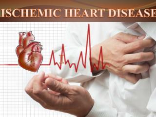
Heart Ischemic Heart Disease • MI = Clinical • Silent MI = DM, Elderly, Cardiac transplantation recipients, • Typical features = Rapid, weak pulse and sweating profusely (diaphoretic), Dyspnea, chest pain • Lab= • Diagnostic • Best markers = Troponins ( T & I), both sensitive and cardio – specific • Next best – CK-MB • Predictive • CRP- >3mg/l – highest risk
Heart Ischemic Heart Disease MI –Complications • In 75% of Patients with MI • Poor prognosis in = elderly, females, DM, old case of MI, Anterior wall infarct – worst, posterior –worse, Inferior wall – best • 1. Arrhythmia = Ventr. Fibrillation – MC arrhythmia lead to sudden death in MI patients, before they reach hospital • 2. pump failure – LVF, cariogenic shock, if >LV wall infarcts, lead to death ( 70% of hospitalized MI patients) • 3.Ventricular rupture = Free or lateral LV wall – MC site, later cause false aneurysm, • 4.True aneurysm = rupture is very rare • 5.Pericarditis = Dressler’s syndrome ( Late MI complication) • 6.Recurrence
Heart Ischemic Heart Disease • Sudden cardiac death = unexpected death in one hour due to cardiac causes with or without clinical symptoms • Cause –Atherosclerosis ( 90%), others (10%) • Romano- Ward syndrome –Long Q-T syndrome ( K+, Na+ channel defects) • Mechanism- Most likely due to arrhythmias ( VF) • Patients – young athletes, with Pul. HTN, IHD • Morphology • Prominent finding – increased heart mass • Vacuolations in Sub – endocardial myocardium
Heart - Ischemic Heart Disease • Chronic IHD =also called ischemic cardiomyopathy • Patients = post heart transplant receipts, previous MI or CABG pts • Cause =compromised ventricular function • Morphology =vacuoles, MyocyteHypertrophy