Download
1 / 44
510 likes | 720 Views
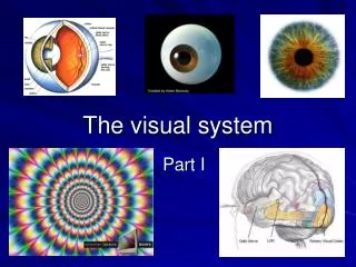
The Human Visual System. Vonikakis Vasilios, Antonios Gasteratos . Democritus University of Thrace 2006 . The Human Visual System. Optic nerve. Visual Cortex V1, V2…. Retina. (ganglion cells). Biological background. light. The eye. Ανθρώπινο Οπτικό Σύστημα. The photoreceptors.

E N D
The Human Visual System Vonikakis Vasilios, Antonios Gasteratos Democritus University of Thrace 2006
The Human Visual System Optic nerve Visual Cortex V1, V2… Retina (ganglion cells) Biological background light
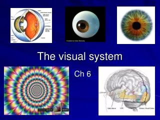
The eye Ανθρώπινο Οπτικό Σύστημα
The photoreceptors • 3 kinds of cones (long, medium, short) – color vision (only in bright light – photopic vision) • Rods – achromatic vision (in dim light – scotopic vision)
Differences from a ccd • Only one layer of photoreceptors • Varying distribution of photoreceptors (Only L and M cones in the fovea, only rods in the periphery) • Different ratios of photoreceptors between individuals (generally L>M>S) • Hexagonal distribution of photoreceptors • No refresh rate – parallel transmission of visual information to the brain
What retina sees Day Night
Basic retinal circuit photoreceptors • Ganglion cells are the only output of from the retina • Digital output with an FM modulation (spikes) Ganglion cell Output
- - + Receptive field • The number of photoreceptors that a ganglion cell “sees” and the kind of the connection • Ganglion cells have antagonistic center-surround receptive field
- - + Center-surround antagonism
- - - - - - - - + + + + Center-surround responses No light light No light light inhibition excitation nothing
Center-surround : facts • Ganglion cells are edge detectors – they respond only to changes and not to uniform areas • By stimulating only the cells that detect differences, the HVS minimizes the number of active neurons • Example: Instead of transmitting a sequence of long numbers e.g. 2003453, 2003453, 2003455, 2003451 it transmits only their differences: 0, 0, +2, -2
Dim light Bright light Center-surround : advantage • White paper in dim light reflects less light (is darker) than the black letters in bright light • The absolute value of reflected light is not important • By responding only to differences, ganglion cells prevent the white paper from being perceived as black
Red-Green oponency Kinds of Ganglion cells Bcenter- (R+G)surround Blue-Yellow oponency Gcenter- Rsurround Rcenter- Gsurround (R+G+B)center - (R+G+B)surround Achromatic opponency Photoreceptor mosaic Biological background
Midget ganglion cells Biological background
Midget ganglion cells • Midget ganglion multiplex 2 signals • Red-Green chromatic opponency • Achromatic high acuity (1 cone = 1 center of the receptive field)
2 Parasol ganglion cells • Parasol ganglion cells are: • Achromatic • Have 3 times greater receptive filed • Respond better to movement
2 Bistratified ganglion cells • Bistratified ganglion cells: • Carry the Blue – Yellow opponency • Have 3 times greater receptive filed
Retinal output • At least 8 independent and parallel mosaics of ganglion cells outputs scan the photoreceptors and transmit different information to the visual cortex
The primary visual cortex V1 • The visual cortex analyses the retinal output in 3 different and independent maps: • color • motion-depth • orientation of edges
The primary visual cortex V1 • The visual cortex analyses the retinal output in 3 different and independent maps: • color • motion-depth • orientation of edges
In every position of the visual field 4 different midget cells (from the 4 mosaics) are connected in couples Chromatic opponency is canceled (same colors to center and surround). Now only sensitive only to luminance increments Center-surround antagonism is canceled Center-surround antagonism is canceled (they are the same) Chromatic opponency is canceled (same colors to center and surround). Now only sensitive only to luminance decrements Demultiplexing RG in cortex
Cell types • For every position of the visual field there are 8 different cells that detect chromatic and achromatic signals in 2 different scales
Cell outputs Red-Green opponency original Blue-Yellow opponency Achromatic (dark-light)
Double opponent cells • Are formed by combinations of simple center-surround cells • Are excited only by chromatic differences of a very specific color (color edges)
Responses • Double opponent cells respond only to very specific changes between certain hues (color edges) original
Simple Orientation cells • Elongated receptive fields (formed by combinations of center-surround receptive fields) • ~12 different orientations (every 15°) • Detect edges of particular orientations only in a very specific position
Complex Orientation cells • Formed by combinations of simple orientation cells • Detect edges of particular orientation anywhere in their receptive field
Orientation cells • At every position of the visual field there are all possible orientations of an edge • Every edge excites a particular orientation cell in a particular position of the visual cortex
Hypercolumns • For every position of the visual field, all cells are grouped into hyper columns • Every hypercolumn is a complete and independent feature detector for a very small part of the visual field • Every hypercolumn contains color cells, orientation cells, disparity cells, motion cells
Hypercolumns • Competition exists between cells of the same hypercolumn and between hypercolumns
Association field Connection of orientation cells • Orientation cells prefer to be connected with others that favor the smooth continuity of contours Biological background
Salient contours • Smooth combinations emerge from the group of orientation cells • This is the first step for contour perception
Contour integration • More complex cells code certain combinations of salient orientation cells
Feature binding • All the features (contours, colors, texture, depth) are being bind in one perception • Binding is described by the Gestalt rules e.g.common fate rule, proximity rule, similarity ruleetc.
Filling-in the features • There is a tendency to spatially diffuse strong signals over the weak ones • This way, regions that do not have a strong feature ‘get’ one from a nearby region that has a strong one • Edges act like barriers that stop the diffusions of the signals • There is filling-in for: • Texture • Color • Disparity
shape color texture motion Binding to one percept Object space binding
Cell(s) for every object • Finally there is one cell (or one population of cells) that respond only to a very specific object • Every perception of an object (either vision triggered or mind triggered) activates these cells • This ‘databank’ of cells is located at the inferior temporal cortex
Inferior temporal cortex • Inferior temporal cortex has columnar organization • Many aspects of an object are stored in neighboring columns • Similar objects are stored in neighboring rows
Inferior temporal cortex • Every object is stored in the object space in many rotated versions • …but we are trained only to the versions we usually see…