Download
1 / 33
330 likes | 336 Views
Learn about the structure and function of the cell membrane and how it maintains homeostasis. Explore the processes of diffusion, osmosis, and active transport. Understand how concentration gradients affect the movement of solutes. Real-life scenarios and diagrams help visualize these concepts.

E N D
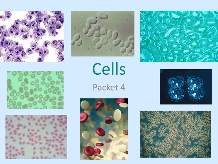
Cells Packet 4
Notes: Cell Membrane Structure PHOSPHOLIPID BILAYER • The cell or plasma membrane is also known as the ________________________________ since it has 2 layers. • It is known as a ______________ ____________ since it is made up of many parts and is not rigid and moves easily. • The main function of the cell membrane is to maintain homeostasis by controlling ___________________________ • The cell membrane doesn’t let everything through. It is _________________________________and only lets certain things in and out. FLUID MOSAIC MODEL WHAT GOES IN AND OUT SELECTIVELY PERMEABLE
Cell Membrane Diagram Video – Cell membrane CHOLESTEROL INTEGRAL PROTEINS CARB CHAIN PHOSPHATE HEAD FATTY ACID TAILS PHOSPHOLIPID PERIPHERAL PROTEIN
Polar, hydrophilic heads Nonpolar hydrophobic fatty acid tails Keep the membrane ____________ FLUID
TRANSPORT RECEIVE SIGNALS IDENTIFY
Concentration Gradient • Differences in concentration on either side of a membrane.
Hill Diagram • Active Transport • Goes against the concentration gradient (low to high) • – needs energy (ATP) • P = Pump – solute through a protein • E = Endocytosis – move into cell • E = Exocytosis – move out of cell High Low High • Passive Transport • Goes with the concentration gradient (high to low) • no energy • D = Diffusion – solute moves (Ex: salt) • O = Osmosis – water moves • F = FacilitatedDiffusion – solute moves through protein Low
DIFFUSION – dots such as salt are moving. OSMOSIS - dots can’t move so water moves to dilute the dots FACILITATED DIFFUSION – dots are too big or polar so need to go through a protein Diffusion Osmosis
water water
Which way will things move? Given the pictures below, draw arrows in the correct direction to show what will move. You need 4 different colors to make your key: Diffusion – RedFacilitated diffusion – Green Osmosis – BluePump (active transport) – Purple The cell is sitting in a _______________________ solution. The cell is sitting in a _______________________ solution. HYPERTONIC HYPOTONIC The cell will _____________________ if ___________________ occurs. The cell will _____________________ if ___________________ occurs. GROW SHRINK OSMOSIS OSMOSIS
Real life scenarios – What will happen??? A saltwater fish is placed into fresh water. What will happen to the cells of this fish? A freshwater fish is placed into a salt water tank. What will happen to the cells of this fish? When it is really humid, doors and windows tend to stick. The higher the humidity the more water there is in the air. Explain why the doors stick? Water will move in and the fish’s cells will grow. Water will move out and the fish’s cells will shrink. Water from the air moves into the wood of the door causing them to swell so they are harder to close.
Explain the following picture. Salt on a slug will cause water to move out of the slug to dilute the salt on the outside of the body. The slug will dehydrate and die.
Hypo = less Hyper = more Iso = same Understanding Osmosis & Diffusion (Passive Transport) When answering the questions, consider the following information: • The oval in each diagram below represents a cell. • The black line around the oval is the cell membrane. • The space between the dots represents the water (solvent) that the solute is dissolved in. • The solute (dots) AND solvent (water) is small enough to pass across the cell membrane. Cell #1 Cell #2 Cell #3
CONCENTRATION DIFFERENCES: • The solution outside cell #1 has a/an (higher; LOWER; equal) concentration of solute compared to the solution inside the cell. • The solution outside cell #1 is (hypertonic; HYPOTONIC; isotonic) to the solution inside the cell. • The inside of cell #1 is (HYPERTONIC; hypotonic; isotonic) to the solution surrounding it. • The solution outside cell #2 has a/an (HIGHER; lower; equal) concentration of solute compared to the solution inside the cell. • The solution outside cell #2 is (HYPERTONIC; hypotonic; isotonic) to the solution inside the cell. • The inside of cell #2 is (hypertonic; HYPOTONIC; isotonic) to the solution surrounding it. • The solution outside cell #3 has a/an (higher; lower; EQUAL) concentration of solute compared to the solution inside the cell. • The solution outside cell #3 is (hypertonic; hypotonic; ISOTONIC) to the solution inside the cell.
DIFFUSION: • If diffusion was to occur to cell #1, in which direction would most of the solute be moving? (into /OUT OF) the cell. • If diffusion was to occur to cell #2, in which direction would most of the solute be moving? (INTO /out of) the cell. • Describe what happens to the movement of solute for cell #3. THE SOLUTE WOULD MOVE IN AND OUT. • Due to the process of diffusion, the solute or dissolved material tries to move from an area of higher concentration into an area of lower concentration (someplace where it can spread out more). According to this statement, which of the above cells would lose the most solute due to diffusion? (CELL #1, CELL #2, CELL #3)
OSMOSIS: • If osmosis was to occur in cell #1, which direction would most of the water be moving? (INTO /out of) the cell. • Cell #1 should have (lost ; GAINED; stayed the same) mass. • If osmosis was to occur in cell #2, which direction would most of the water be moving? (into /OUT OF) the cell. • Cell #2 should have (LOST ; gained; stayed the same) mass. • If osmosis was to occur in cell #3, which direction would most of the water be moving? (INTO & OUT OF) the cell. • Cell #3 should have (lost ; gained; STAYED THE SAME) mass.
WHAT DOES THIS MEAN… • Since an animal cell lacks a cell wall, it is important that it be surrounded by a/an (hypertonic; hypotonic; ISOTONIC) solution, so that it doesnot shrink & shrivel up or swell & rupture due to the effects of osmosis. • If red blood cell is surrounded by a hypotonic solution, then the cell would most likely (shrink, SWELL or stay the same size). • When plant cells are full of water, the pressure within the cell pushes out onto the cell wall, thus allowing the cell to become more rigid (has turgor pressure). Since this is a good thing for them, plant cells should be surrounded by a/an (hypertonic; HYPOTONIC; isotonic) solution.
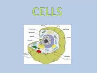
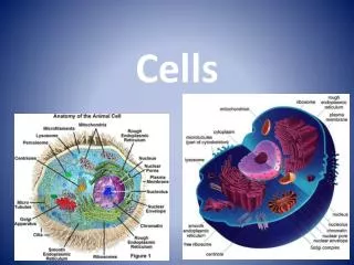
Cytoplasmic Organelles An ORGANELLE is a tiny structure that performs a specialized function (or job) in the cell. Nucleolus DNA NUCLEUS NO YES YES Nuclear Envelope CELL MEMBRANE YES YES YES YES YES NO CELL WALL Cell Membrane RIBOSOME YES YES YES Cell Wall
ENDOPLASMIC RETICULUM (E.R) NO YES YES GOLGI APPARTUS (BODIES) NO YES YES LYSOSOMES NO NO YES NO YES NO CHLOROPLAST GLUCOSE PHOTOSYNTHESIS
ATP MITOCHONDRIA NO YES YES CELLULAR RESPIRATION Plant Cell NO YES YES (Large (Small) Central) Vacuole VACUOLE Nucleus YES YES YES CYTOSKELETON
FLAGELLUM YES NO YES Flagellum CILIA NO NO YES CENTRIOLES NO NO YES Pathway for Proteins: proteins made at ribosome proteins travel thru E.R. Proteins modified/ sorted/shipped out of Golgi
Notes: Prokaryotic versus Eukaryotic Cells Cell Theory • All living things are made up of one or more cells • Cells are the basic unit of structure and function of an organism. • All cells come from pre-existing cells. All cells can be divided into one of two categories based upon their complexity: • Prokaryotic Cells are very • Donot have a __________(the part that contains the DNA) • Do not have membrane bound • Usually small and _______________(meaning they are made up of a single cell) • Example: SMALL NUCLEUS ORGANELLES UNICELLULAR BACTERIA
Eukaryotic Cells are • Have a • Have membrane bound • Can be both unicellular and • Example: Cells of • Structures that are common to all cells are: LARGER NUCLEUS ORGANELLES MULTICELLULAR PLANTS & ANIMALS DNACELL MEMBRANE CYTOPLASMRIBOSOMES
Complete the table by checking the correct column for each statement:
How did the first Eukaryotic cells come about? INSIDE RELATIONSHIP PROKARYOTES Endosymbiotic Theory – endo means ____________________________, symbiotic means ____________________________ so…… • Smaller __________________________ entered inside larger prokaryotes. • The smaller prokaryote(s) got ___________________________________. • The larger prokaryote got _____________________________________. • This smaller cell was really good at producing _____________ and came to be known as the _________________________. • Eventually a 2nd small prokaryotic cell was engulfed by the larger cell… • This cell was really good at absorbing solar energy and using it to make food for itself and came to be known as the _________________. PROTECTION ENERGY ATP MITOCHONDRIA CHLOROPLAST
Support for the endosymbiotic theory: REPRODUCE • Unlike other organelles, mitochondria & chloroplasts both ___________________ independently of the cell. • Both mitochondria & chloroplasts contain their own __________ • Both have two ___________________around them. DNA MEMBRANES AUTOGENY _____________________________ Theory – organelles formed when the plasma membrane folded inward producing membranes inside of the cell. Some of the infolds broke off forming various organelles. Endosymbiosis Animation
Cell Specialization • Why are cells specialized? • So different cells can perform different functions. • What are stem cells? – • Cells that can become anything. They have not differentiated or become specialized yet.
Microscopes OBJECTIVES and OCULAR (EYEPIECE) Types of Microscopes • Compound Light Microscope: • Uses two lenses: • Used to magnify and show detail of a specimen (one that light can pass through). • Image is as a result of the lenses that are used. • Can magnify up to 1500-2000 times (The ones we have only magnify up to times). • The equation below is used to calculate total magnification: • Total magnification = magnification of X magnification of • Ex: If the eyepiece magnifies the image 10x and the low power objective magnifies the image 10x, then the total magnification of the image would be: TRANSLUCENT INVERTED 400 OBJECTIVElensOCULARlens 10 x 10 = 100
Microscopes Continued FIELD OF VIEW SMALLER • - This is the visible area seen through the microscope when the specimen is in focus. • The greater the magnification, the ___________ the field of view will be. LOW POWER MEDIUM POWER HIGH POWER DISSECTING 2. Stereomicroscope (also known as ________________________Microscope). Useful when viewing objects that are too ____________________or too ___________for the compound light microscope. Unlike the compound light microscope, the images seen are not inverted. This is because you are using _______________eyepieces (binocular scope). Can only magnify up to _______ - _______times. LARGE THICK TWO 4X – 50X
ELECTRON MICROSCOPES SEM SURFACE 100,000 3. Scanning Electron Microscopes – _______* • Shows the ________________only • Can magnify up to __________times TEM • 4. Transmission Electron Microscope – ___________* • Electrons pass through the object - allows you to “see”_______ • Can magnify up to ___________times • ** A big problem of the SEM and TEM is that the specimen must be ______________________ in order to view in with one of these microscopes. INSIDE 200,000 DEAD