Download
1 / 38
380 likes | 391 Views
Amino Acids: Disposal of Nitrogen. Overview Unlike fats and carbohydrates, amino acids are not stored by the body, That is no protein exists whose sole function is to maintain a supply of amino acids for future use.

E N D
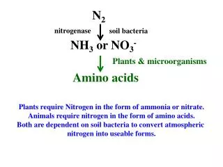
Overview • Unlike fats and carbohydrates, amino acids are not stored by the body, • That is no protein exists whose sole function is to maintain a supply of amino acids for future use. • Therefore, amino acids must be obtained from the diet, synthesized de novo, or produced from normal protein degradation.
Any amino acids in excess of the biosynthetic needs of the cell are rapidly degraded. • The first phase of catabolism involves the removal of the α-amino groups (usually by transamination and subsequent oxidative deamination), • Forming ammonia and the corresponding α-keto acid—the “carbon skeletons” of amino acids. A portion of the free ammonia is excreted in the urine, but most is used in the synthesis of urea
Synthesis of ammonia is quantitatively the most important route for disposing of nitrogen from the body. • In the second phase of amino acid catabolism, the carbon skeletons of the α-ketoacids are converted to common intermediates of energy producing, metabolic pathways. • These compounds can be metabolized to CO2 and water, glucose, fatty acids, or ketone bodies by the central pathways of metabolism. • Amino acid catabolism is part of the larger process of the metabolism of proteins.
Protein metabolism • Protein metabolism or proteinolysis • Denotes the various biochemical processes responsible for the synthesis of proteins and amino acids • the breakdown of proteins (and other large molecules, too) by catabolism
Pepsin nonspecific maximally active at low pH of the stomach. • Proteolytic enzymes of the pancreas in the intestinal lumen display a wide array of specificity. Aminopeptidases digest proteins from the amino-terminal end. The Digestion and Absorption of Dietary Proteins
Some proteins are very stable, while others are short lived. • Altering the amounts of proteins important in metabolic regulation can rapidly change metabolic patterns. • Cells have mechanisms for detecting and removing damaged proteins. • A significant proportion of newly synthesized protein molecules are defective because of errors in translation. • Other proteins may undergo oxidative damage or be altered in other ways with the passage of time. Cellular Proteins Are Degraded at Different Rates
Ubiquitin Tags Proteins for Destruction • How can a cell distinguish proteins that are meant for degradation? • Ubiquitin, a small (8.5-kd) protein present in all eukaryotic cells, is the tag that marks proteins for destruction.
The c-terminal glycine residue of ubiquitin (Ub) becomes covalently attached to the e-amino groups of several lysine residues on a protein destined to be degraded. • The energy for the formation of these isopeptidebonds (iso because e- rather than a-amino groups are targeted) comes from ATP hydrolysis.
Three enzymes participate in the attachment of ubiquitin to each protein: • ubiquitin-activating enzyme, or E1 • ubiquitin-conjugating enzyme, or E2 • ubiquitin-protein ligase, or E3.
Chains of ubiquitin can be generated by the linkage of the e-amino group of lysine residue 48 of one ubiquitin molecule to the terminal carboxylate of another. • Chains of four or more ubiquitin molecules are particularly effective in signaling degradation
The half-life of a cytosolic protein is determined to a large extent by its amino-terminal residue “the N-terminal rule”. • In yeast: if N terminus is methionine half-life > 20 hours, whereas if N terminus is arginine half-life ≈ 2 minutes. • A highly destabilizing N-terminal residue such as arginine or leucine favors rapid ubiquitination, whereas a stabilizing residue such as methionine or proline does not. • E3 enzymes are the readers of N-terminal residues. What determines whether a protein becomes ubiquitinated?
Cyclin destruction boxes are amino acid sequences that mark cell-cycle proteins for destruction. • Proteins rich inproline, glutamic acid, serine, and threonine (PEST sequences).
A large protease complex called the proteasome or the 26S proteasome digests the ubiquitinated proteins. • In eukaryotes, they are located in the nucleus and the cytoplasm. • The degradation process yields peptides of about 7-8 amino acids long, then further degraded into amino acids and used in synthesizing new proteins. • This ATP-drivenmultisubunit protease spares ubiquitin, which is then recycled. The Proteasome Digests the Ubiquitin-Tagged Proteins
P P E3 P P Ub NF-kB NF-kB I-kB P P Ub Ub P Ub P Ub proteosome Example: E3 Inflammation initiates the expression of a number of the genes that take part in this response Protein Degradation Can Be Used to Regulate Biological Function
Digested proteins Amino Acids Degradation in the liver NH4+ a-ketoacids The amino group must be removed, as there are no nitrogenous compounds in energy-transduction pathways enter the metabolic mainstream as precursors to glucose or citric acid cycle intermediates
The fate of the a-amino group • The a-amino group of many aas is transferred to a-ketoglutarate to form glutamate. • Glutamate is then oxidatively deaminated to yield ammonium ion (NH4+).
Aminotransferases (transaminases) catalyze the transfer of an a-amino group from an a-amino acid to an a-keto acid.
Aspartate aminotransferase: • Alanine aminotransferase: • These transamination reactions are reversible and can thus be used to synthesize amino acids from a-ketoacids, Example:
The nitrogen atom that is transferred to a-ketoglutarate in the transamination reaction is converted into free ammonium ion by oxidative deamination. • This reaction is catalyzed by glutamate dehydrogenase. • This enzyme is unusual in being able to utilize eitherNAD+ or NADP+ at least in some species. • The reaction proceeds by dehydrogenation of the C-N bond, followed by hydrolysis of the resulting Schiff base.
Glutamate dehydrogenase and other enzymes required for the production of urea are located in mitochondria. • This compartmentalization sequesters free ammonia, which is toxic. • In most terrestrial vertebrates, NH4+ is converted into urea, which is excreted.
Pyridoxal Phosphate Forms Schiff-Base Intermediates in Aminotransferases • All aminotransferases contain the prosthetic group pyridoxal phosphate (PLP), which is derived from pyridoxine (vitamin B6).
The most important functional group allows PLP to form covalent Schiff-base intermediates with amino acid substrates Pyridoxal phosphate derivatives can form stable tautomeric forms a pyridine ring that is slightly basic A phenolic hydroxyl group that is slightly acidic
The aldehyde group of PLP usually forms a Schiff-base linkage with the e-amino group of a specific lysine residue of the enzyme. • The a-amino group of the amino acid substrate displaces the e-amino group of the active-site lysine residue.
Some of the NH4+ formed in the breakdown of amino acids is consumed in the biosynthesis of nitrogen compounds. • In most terrestrial vertebrates, the excess NH4+ is converted intourea and then excreted. • The urea: • One nitrogen atom is transferred from aspartate. • The other nitrogen atom is derived directly from free NH4+ . • The carbon atom comes from HCO3-. The Urea Cycle
Formation of Carbamoyl Phosphate: catalyzed by carbamoyl phosphate synthetase. • The consumption of two molecules ofATP makes the synthesis essentially irreversible. The Urea Cycle Reactions
Carbamoyl is transferred to ornithine to form citrulline. • The reaction is catalyzed by ornithine transcarbamoylase. • Ornithine and citrulline are amino acids, but they are not used as building blocks of proteins.
Citrulline is transported to the cytoplasm where it condenses with aspartate to form argininosuccinate • The reaction is catalyzed by argininosuccinatesynthetase. • The reaction is driven by the cleavage ofATP into AMP and PPi, and by the subsequent hydrolysis ofPPi.
Argininosuccinase cleaves argininosuccinate into arginine and fumarate. • Thus, the carbon skeleton of aspartate is preserved in the form of fumarate.
Arginine is hydrolyzed to generate urea and ornithine in a reaction catalyzed by arginase. • Ornithine is then transported back into the mitochondrion to begin another cycle.
Mitochondrial reactions: • The formation of NH4+ by glutamate dehydrogenase. • Its incorporation into carbamoyl phosphate • Synthesis of citrulline • Cytosolic reactions: • The next three reactions of the urea cycle, which lead to the formation of urea, take place in the cytosol.
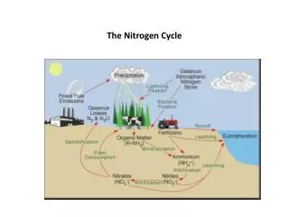
From amino acids: Many tissues, but particularly the liver, form ammonia from amino acids by transdeamination—the linking of aminotransferase and glutamate dehydrogenase reactions. • From glutamine: The kidneys form ammonia from glutamine by the actions of renal glutaminase and glutamate dehydrogenase. Most of this ammonia is excreted into the urine as NH4+, which provides an important mechanism for maintaining the body's acid-base balance. Sources of ammonia
From bacterial action in the intestine: Ammonia is formed from urea by the action of bacterial ureasein the lumen of the intestine. This ammonia is absorbed from the intestine by way of the portal vein and is almost quantitatively removed by the liver via conversion to urea. • From amines: Amines obtained from the diet, and monoamines that serve as hormones or neurotransmitters, give rise to ammonia by the action of amine oxidase. • From purines and pyrimidines: In the catabolism of purines and pyrimidines, amino groups attached to the rings are released as ammonia.
The capacity of the hepatic urea cycle exceeds the normal rates of ammonia generation, and the levels of serum ammonia are normally low (5–50 µmol/L). • However, when liver function is compromised, due either to genetic defects of the urea cycle, or liver disease, blood levels can rise above 1,000 µmol/L. • Such hyperammonemia is a medical emergency, because ammonia has a direct neurotoxic effect on the CNS. • For example, elevated concentrations of ammonia in the blood cause the symptoms of ammonia intoxication, which include tremors, slurring of speech, somnolence, vomiting, cerebral edema, and blurring of vision. At high concentrations, ammonia can cause coma and death. Hyperammonemia
The two major types of hyperammonemia: • Acquired hyperammonemia: Liver disease is a common cause of hyperammonemia in adults. It may be a result of an acute process (for example, viral hepatitis, ischemia, or hepatotoxins). Cirrhosis of the liver caused by alcoholism, hepatitis, or biliary obstruction may result in formation of collateral circulation around the liver. As a result, portal blood is shunted directly into the systemic circulation and does not have access to the liver. The detoxification of ammonia (that is, its conversion to urea) is, therefore, severely impaired, leading to elevated levels of circulating ammonia.
2. Hereditary hyperammonemia: • Genetic deficiencies of each of the five enzymes of the urea cycle have been described, with an overall prevalence estimated to be 1:30,000 live births. • Ornithinetranscarbamoylase deficiency, which is X-linked, is the most common of these disorders, predominantly affecting males, although female carriers may become symptomatic. • The failure to synthesize urea leads to hyperammonemia during the first weeks following birth.
All inherited deficiencies of the urea cycle enzymes typically result in mental retardation. • Treatment includes limiting protein in the diet, • and administering compounds that bind covalently to amino acids, producing nitrogen-containing molecules that are excreted in the urine. • For example, phenylbutyrate given orally is converted to phenylacetate. This condenses with glutamine to form phenylacetyl-glutamine, which is excreted.