Download
1 / 34
380 likes | 720 Views
DNA polymerase III. Enzyme used during replication Multisubunit protein High processivity. DNA polymerase III = DNA Pol III. Discovered in extracts of polA - cells, i.e. lacking DNA Pol I DNA Pol III is the replicative polymerase

E N D
DNA polymerase III Enzyme used during replication Multisubunit protein High processivity
DNA polymerase III = DNA Pol III • Discovered in extracts of polA- cells, i.e. lacking DNA Pol I • DNA Pol III is the replicative polymerase • Loss-of-function mutations in the genes encoding its subunits block DNA replication (dna mutants) • Highly processive • Multiple subunits • Also discovered DNA Pol II in polA- extracts (role in DNA repair)
DNA Pol III: Low abundance but high processivity Comparison Pol IPol III corePol III holo molecules per cell 400 40 10 nts polymerized min-1 (molecule enz)-1600 9000 42,000 processivity [nts polymerized per initiation] 3-188 10 >105 5' to 3' polymerase + + + 3' to 5' exo, proofreading + + + 5' to 3' exo + - -
Processivity • Amount of polymerization catalyzed by an enzyme each time it binds to a template. • Measured in nucleotides polymerized per initiation • High processivity of DNA Pol III results from activities of non-polymerase subunits
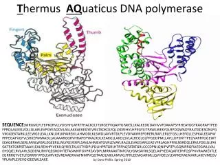
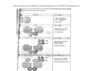
Subunits of DNA Pol III and functions Functional Mass component Subunit (kDa) Gene Activity Core polymerase a 129.9 polC=dnaE 5' to 3' polymerase e 27.5 dnaQ=mutD 3'-5' exonuclease q 8.6 Stimulates e exonuclease Linker protein t 71.1 dnaX Dimerizes cores Clamp loader g 47.5 dnaX Binds ATP (aka g complex) d 38.7 Binds to b (ATPase) d' 36.9 Binds to g and b c 16.6 Binds to SSB y 15.2 Binds to c and g Sliding clamp b 40.6 dnaN Processivity factor
Processivity factor beta2: Sliding clamp • The b subunit forms a homodimer. • The structure of this homodimer is a ring. • The ring encloses DNA, thereby clamping the DNA Pol III holoenzyme to the template. • An enzyme that is clamped on cannot come off easily, and thus will be highly processive.
Gamma complex: Clamp loader/unloader • The g complex (g2dd'cy) loads the b dimer clamp onto a primer-template. • Bind the clamp (b dimer) onto the loader (g complex): need ATP • Exchange the clamp from the loader to the core: need ATP hydrolysis • Unload the clamp when polymerase reaches a previously synthesized Okazaki fragment: need ATP • The ATP-bound form of the g complex can bind the clamp • The ADP-bound form releases the clamp
Model for gamma complex loading beta clamp Jeruzalmi, O’Donnell and Kuriyan (2001) Cell 106: 429-441
Asymmetric dimer of DNA PolIII: simultaneous replication of both strands of DNA • The 2 catalytic cores of DNA Pol III are joined by the tau subunits to make an asymmetric dimer. • Model: one holoenzyme synthesizes both strands at a replication fork. • One core synthesizes the leading strand • Other synthesizes the lagging strand. • If the template for lagging strand synthesis is looped around the enzyme, then both strands are synthesized in the direction of fork movement.
Eukaryotic replicative DNA polymerases • Nuclear DNA replication: • a: primase plus low processivity polymerase • d: both leading and lagging strand synthesis • e: may be used in lagging strand synthesis
Similarities between bacterial and eukaryotic replication machinery FunctionE. coli Pol IIIEukaryotic Leading and lagging asymmetric polymerase d strand synthesis dimer Sliding clamp b subunit PCNA Clamp loader g-complex RFC Primase DnaG polymerase a Single strand binding SSB RFA Swivel Gyrase (Topo II) Topo I or II (Maintain DNA topology)
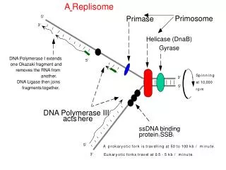
Enzymes other than polymerases needed for replication Helicases Ligases Primosome
DNA helicases • Unwind the DNA duplex as the replication fork moves. • Use ATP: Hydrolyze 2 ATPs to 2ADP+2Pi for every base pair that is unwound. • In addition, helicases move along single stranded DNA with a specific polarity; referred to as tracking.
Assay for helicase movement, #1 B A DnaB + ATP A B + Displacement of A shows that DnaB moved 5’ to 3’ along the single-stranded DNA.
Assay for helicase movement, #2 B A PriA + ATP B A + Displacement of B shows that PriA moved 3’ to 5’ along the single-stranded DNA.
Single-stranded binding protein (SSB) • Encoded by the ssb gene in E. coli. • Loss-of-function mutants in ssb have a quick-stop phenotype for DNA synthesis. They are also defective in repair and recombination. • Binds cooperatively to single-stranded DNA to prevent reannealing to the complementary strand. • SSB is a homo-tetramer, monomer is 74 kDa • Eukaryotic RFA (analog to SSB) is aheterotrimer.
Topoisomerases • Topoisomerase I: relaxes DNA • Transient break in one strand of duplex DNA • E. coli: nicking-closing enzyme • Calf thymus Topo I • Topoisomerase II: introduces negative superhelical turns • Breaks both strands of the DNA and passes another part of the duplex DNA through the break; then reseals the break. • Uses energy of ATP hydrolysis • E. coli: gyrase
DNA ligases • Join together the Okazaki fragments during lagging strand synthesis • Tie together a nick
Primase • Synthesizes short oligonucleotides from which DNA polymerases can begin synthesis. • Combination of ribonucleotides and deoxyribonucleotides • Does not itself require a primer. • E. coli primase is DnaG, 60 kDa • Acts within a large primosome.
Primers made by DnaG • Primers can be as short as 6 nt, as long as 60 nt. • Can substitute dNTPs for rNTPs in all except 1st and 2nd positions • Make hybrid primers with dNMPs and rNMPs interspersed. • Primase binds to CTG • T serves as template for 1st nucleotide of primer.
Primosome has many proteins Pre-priming complex: Protein gene function PriA priA helicase, 3' to 5' movement, site recognition PriB priB PriC priC DnaT dnaT needed to add DnaB-DnaC complex to preprimosome DnaC dnaC forms complex with DnaB DnaB dnaB helicase, 5' to 3' movement, is a hexamer DNA dependent ATPase. Primase = DnaG
Assay for assembly and migration of the primosome Convert single stranded (ss) fX174 to duplex, replicative form (RF)
Activities of DnaB and PriA in replisome “Sewing machine” model
E. coli: 50,000 bp per min Plants and animals: 1000 to 3000 bp per min Need many origins for replication of large genomes Rate of fork movement