Download
1 / 89
920 likes | 1.53k Views
Peptic Ulcer Disease (PUD). DEFINITION. Peptic ulcer disease (PUD) refers to a group of ulcerative disorders of the upper GI tract that require acid and pepsin for their formation. Ulcers differ from gastritis and erosions in that they extend deeper into the muscularis mucosa .

E N D
DEFINITION • Peptic ulcer disease (PUD) refers to a group of ulcerative disorders of the upper GI tract that require acid and pepsin for their formation. • Ulcers differ from gastritis and erosions in that they extend deeper into the muscularismucosa. As many as 70–90% of such ulcers are associated with Helicobacter pylori, a rod-shaped bacterium that lives in the acidic environment of the stomach; however, only 40% of those cases go to a doctor. Ulcers can also be caused or worsened by drugs such as aspirin, ibuprofen, and other NSAIDs. • The three common forms of peptic ulcers include: • Helicobacter pylori (HP)– associated ulcers, • Non-steroidal antiinflammatory drug (NSAID)–induced ulcers, • And stress-related mucosal damage (also called stress ulcers).
The parietal cell is regulated by neural, hormonal, and paracrine pathways. • Activation of vagal parasympathetic preganglionic outflow to the stomach acts in three ways to stimulate gastric acid secretion. • There is direct neural innervation and activation of the parietal cell via release of acetylcholine (ACh) from enteric neurons, which acts on the parietal cell via muscarinic receptors. • In addition, neural activation of the ECL cell stimulates the release of histamine, which acts via a paracrine pathway to stimulate the parietal cell. • Finally, G cells located in gastric glands in the gastric antrum are activated by the release of gastrin-releasing peptide from enteric neurons, which acts on the G cell to stimulate the release of gastrin. Gastrin thereafter acts via a humoral pathway to stimulate the parietal cell.
Components involved in providing gastroduodenal mucosal defense and repair HCO3 HCO3 HCO3 HCO3
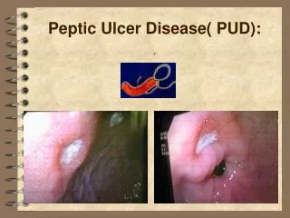
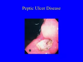
What Causes Peptic Ulcer Disease Helicobacter Pylori (H. pylori) • Most ulcers are the result of infection with H. pylori • Not all of those infected with H. pylori develop ulcers • H. pylori MAY result in a weakening of the mucosal defense systems, allowing for development of ulcer subsequent to acid/pepsin aggression;
Pathogenesis In normal acid/pepsin attack is balanced by mucosal defences Increased attack by hyperacidity Weakened mucosal defence – the major factor (H. pylori related)
Gram negative bacterium Figure 28.3 (still) Chapter 28 MENU >
H. pylori produces large amounts of the enzyme urease, molecules of which are localized inside and outside of the bacterium. Urease breaks down urea (which is normally secreted into the stomach) to carbon dioxide and ammonia (which neutralizes gastric acid). • The survival of H. pylori in the acidic stomach is dependent on urease, and it would eventually die without the enzyme. • The ammonia that is produced is toxic to the epithelial cells, and, along with the other products of H. pylori—including protease, vacuolatingcytotoxin A (VacA), and certain phospholipases—damages those cells. • Thus • 1. H. pylori penetrate the mucus layer of host stomach and adhere the surface of gastric mucosal epithelial cells. • 2. produce ammonia from urea by the urease, and the ammonia netralize the gastric acid to escape from elimination. • 3. prolifirate, migrate, and finally form the infectious focus. • 4. The gastric ulcerization is developed by destruction of mucosa, inflammation and mucosal cell death.
What Causes Peptic Ulcer Disease • NonSteroidalAntiInflammatory Drugs (NSAIDs) Long term use of nonsteroidal anti-inflammatory drugs. NSAIDs block COX enzymes and decrease prostaglandins (PGs). • Gastrinoma (Zollinger-Ellison Syndrome) Tumors of the duodenum or pancreas and secrete abnormally high amounts of gastrin which stimulates gastric acid. • Stress ulcers Result of physical trauma
PATHOPHYSIOLOGY • The pathogenesis of duodenal ulcers (DU) and gastric ulcers (GU) is multifactorialand most likely reflects a combination of pathophysiologic abnormalities and environmental as well as genetic factors. • Most peptic ulcers occur in the presence of acid and pepsin when HP, NSAIDs, or other factors disrupt normal mucosal defense and healing mechanisms. • Acid is an independent factor that contributes to disruption of mucosal integrity. Increased acid secretion has been observed in patients with DU and may result from HP infection. Patients with GU usually have normal or reduced rates of acid secretion. • Alterationsin mucosal defense induced by HP or NSAIDs are the most important cofactors in peptic ulcer formation.
PATHOPHYSIOLOGY (Cont.) • Mucosal defense and repair mechanisms include mucus and bicarbonate secretion, intrinsic epithelial cell defense, and mucosal blood flow. • Maintenance of mucosal integrity and repair is mediated by endogenous prostaglandinproduction. • HP infection causes gastritis in all infected individuals and is causally linked to PUD. However, only about 20% of infected persons develop symptomatic PUD. • Most non-NSAID ulcers are infected with HP, and HP eradication markedly decreases ulcer recurrence. HP may cause ulcers by direct mucosal damage, altering the immune/inflammatory response, and by hypergastrinemia leading to increased acid secretion.
PATHOPHYSIOLOGY (Cont.) • Nonselective NSAIDs (including aspirin) cause gastric mucosal damageby two mechanisms: • (1) A direct or topical irritation of the gastric epithelium, and • (2) Systemic inhibition of the cyclooxygenase-1 (COX-1) enzyme, which results in decreased synthesis of protective prostaglandins. • Use of corticosteroids alone does not increase the risk of ulcer or complications, but ulcer risk is doubled in corticosteroid users taking NSAIDs concurrently.
Other Factors: • Epidemiologic evidence links cigarette smoking to PUD, impaired ulcer healing, and ulcer-related GI complications. • The risk is proportional to the amount smoked per day. • Although clinical observation suggests that ulcer patients are adversely affected by stressful life events, controlled studies have failed to document a cause-and-effect relationship. • Coffee, tea, cola beverages, beer, milk, and spices may cause dyspepsia but do not increase PUD risk. • Ethanol ingestion in high concentrations is associated with acute gastric mucosal damage and upper GI bleeding but is not clearly the cause of ulcers.
CLINICAL PRESENTATION • Abdominal pain is the most frequent symptom of PUD. The pain is often epigastricand described as burning but can present as vague discomfort, abdominal fullness, or cramping. • A typical nocturnal pain may awaken patients from sleep, especially between 12 AM and 3 AM. • Pain from DU often occurs 1 to 3 hours after meals and is usually relieved by food, whereas food may precipitate or accentuate ulcer pain in GU. • Antacids provide rapid pain relief in most ulcer patients. • Heartburn, belching, and bloating often accompany the pain. • Nausea, vomiting, and anorexia are more common in GU than DU.
CLINICAL PRESENTATION • The severity of symptoms varies from patient to patient and may be seasonal, occurring more frequently in the spring or fall. • Pain does not always correlate with the presence of an ulcer. • Asymptomatic patients may have an ulcer at endoscopy, and patients may have persistent symptoms even with endoscopically proven healed ulcers. • Many patients (especially older adults) with NSAID-induced, ulcer-related complications have no prior abdominal symptoms.
Complications: • Complications of ulcers caused by HP and NSAIDs include: • Upper GI bleeding, • Perforation into the peritoneal cavity, • Penetration into an adjacent structure (ex., Pancreas, biliary tract, or liver), • And gastric outlet obstruction. • Bleeding may be occult or present as melena or hematemesis. • Perforation is associated with sudden, sharp, severe pain, beginning first in the epigastrium but quickly spreading over the entire abdomen. • Symptoms of gastric outlet obstruction typically occur over several months and include early satiety, bloating, anorexia, nausea, vomiting, and weight loss.
DIAGNOSIS • The physical examination may reveal epigastric tenderness between the umbilicus and the xiphoid process that less commonly radiates to the back. • Routine laboratory tests are not helpful in establishing a diagnosis of uncomplicatedPUD. The hematocrit, hemoglobin, and stool hemocculttests are used to detect bleeding.
Diagnostic Procedures: • The diagnosis of HP infection can be made using endoscopic or nonendoscopic tests. • The tests that require upper endoscopy are invasive, more expensive, uncomfortable, and usually require a mucosal biopsy for histology, culture, or detection of urease activity. • The nonendoscopic tests include serologic antibody detection tests, the urea breath test (UBT), and the stool antigen test. • Serologic tests detect circulating immunoglobulin G directed against HP but are of limited value in evaluating post-treatment eradication. • The Urea Breath Test is based on urease production by HP. • Testing for HP is only recommended if eradication therapy is considered.
Figure 28.2 (still) Chapter 28 MENU >
Urease Ammonia cloud Urease Urease Urease Urease Urease Urease H2O Urea Urease Urease Urease Urease 2CO2 NH3 Urease Urease Urease Urease Urease Urease Type IV secretion system Urease Urease Urease
Diagnostic Procedures: • If endoscopy is not planned, serologic antibody testing is reasonable to determine HP status. The UBT is the preferred nonendoscopic method to verify HP eradication after treatment. • The diagnosis of PUD depends on visualizing the ulcer crater either by upper GI radiography or endoscopy. Radiography may be the preferred initial diagnostic procedure in patients with suspected uncomplicated PUD. • Upper endoscopy should be performed if complications are thought to exist or if an accurate diagnosis is warranted. If a GU is found on radiography, malignancy should be excluded by direct endoscopic visualization and histology.
DESIRED OUTCOME • The goals of treatment are relieving ulcer pain, healing the ulcer, preventing ulcer recurrence, and reducing ulcer-related complications. • In HP positive patients with an active ulcer, a previously documented ulcer, or a history of an ulcer-related complication, the goals are to eradicate the organism, heal the ulcer, and cure the disease with a cost-effective drug regimen.
NON-PHARMACOLOGIC TREATMENT • Patients with PUD should eliminate or reduce psychological stress, cigarette smoking, and the use of nonselective NSAIDs (including aspirin). • If possible, alternative agents such as acetaminophen, a nonacetylated salicylate (e.g., salsalate), or a COX-2 selective inhibitor (celecoxib, rofecoxib) should be used for pain relief. • Although there is no need for a special diet, patients should avoid foods and beverages that cause dyspepsia or exacerbate ulcer symptoms (e.g., spicy foods, caffeine, alcohol).
PHARMACOLOGIC TREATMENT • Eradication of HP is recommended for HP-infected patients with GU, DU, ulcer-related complications, and in some other situations. • Treatment should be effective, well tolerated, easy to comply with, and cost-effective.
Helicobacter eradication Use of multiple drugs prevents development of drug resistance Quadruple Therapy Bismuth Subsalicylate Metronidazole Tetracycline H2 receptor antagonist or PPI More commonly given for 2 weeks Gastric acid suppressors for 6-8 weeks Eradication rate = 90%
0 Strategies for Protecting the Gastric Mucosa from Acid Exposure Mechanisms Example Cimetidine Omeprazole Prostaglandins Muscarinic antagonists Inhibit secretion H+ Prevent contact H+ Sucralfate Bismuth Neutralize acid Antacids H+
H+, K+-ATPase (the proton pump) is the final transport pathway for parietal cell hydrogen ion secretion • H+, K+-ATPase is located on the parietal cells of the gastric mucosa • The pump requires large amounts of energy that is supplied by intracellular ATP; • Inhibition of H+, K+-ATPase blocks HCL into the lumen of the stomach
Proton-Pump inhibitors (PPIs) • These drugs are prodrugs with an acid-resistant enteric coating to protect them from degradation in stomach. • The coating is removed in the alkaline duodenum, and the prodrug, a weak base, is absorbed and transported to the parietal cell where it is converted to the active form forming a stable covalent (irreversible) bond (ATPase needs to be resynthesized to overcome this inhibition which takes 18 hrs).
Omeprazole • H+/ K+-ATPase inhibitor; first of PPI class • Profound reduction of gastric acid - elevates gastric pH significantly (20mg/day for 7days will decrease acid by 95%); • Highly protein bound; Metabolized by CYP2C & CYP3A; plasma half life of 1 to2 hours but long duration of action; • Should be taken just prior to a meal and should NOT be taken with other acid-suppressing agents.
0 Esomeprazole Simply the S-isomer of omeprazole; H+, K+-ATPase inhibitor; Given orally. Pantoprazole H+, K+-ATPase inhibitor; An acid-stable form and can be given by IV
0 Proton Pump Inhibitors (PPI) Generally well tolerated • Hypergastrinemia (can lead to tumor growth in the GI) • Nausea • Headaches, skin rashes
Adverse effects of PPIs Figure 28.7 (still) Chapter 28 MENU >
Histamine H2 Antagonists • Cimetidine • Ranitidine • Famotidine • Nizatidine
Drugs for Acid-Peptic Disorders - Cimetidine • Competitive H2 receptor Antagonist; • inhibits basal acid secretion including nocturnal secretion; • Readily absorbed after oral administration; • Relatively brief duration of action (4-8 hr) • Given on a multiple dosing schedule; • (300-400 mg, 2-4 times daily); • Typical therapy is for 4-8 weeks • .
Figure 28.5 (still) Chapter 28 MENU >
Figure 28.6 (still) Chapter 28 MENU >
Drugs for Acid-Peptic Disorders - Cimetidine • Side effects include inhibition of the microsomal metabolism of other drugs results in higher blood levels and enhancement of their effects • Interactions have been shown with: Diazepam Chlordiazepoxide Theophylline Phenytoin Warfarin Propranolol Meperidine Pentobarbital Lidocaine and many others...
Drugs for Acid-Peptic Disorders - Cimetidine Additional Side effects: • In some patients, cimetidine acts as a nonsteroidalantiandrogen (i.e., interferes with estrogen metabolism). • It results in gynecomastia (swelling of the breasts and soreness of the nipples in males) • Can produce confusion and disorientation in elderly patients; • Diarrhea, rash and miscellaneous other effects in a small number of patients.
Drugs for Acid-Peptic Disorders – Ranitidine, Famotidine, Nizatidine • Same mechanism of action as Cimetidine but a longer duration of action (8 to 12 hrs); • Given orally; can be given less frequently than cimetidine • Less interactions on P450 than Cimetidine.
0 Drugs for Acid-Peptic Disorders - Anticholinergics • General muscarinic receptor antagonists • Dicyclomine Side-effects are typical of anticholinergics such as • Dry mouth • Tachycardia • Blurred vision • Bowel discomfort (constipation) • Difficulty in urination
Inhibits: Acid secretion Gastrin release Pepsin secretion Stimulates: Mucus secretion Bicarbonate secretion Mucosal blood flow 0 Drugs for Acid-Peptic Disorders – Prostaglandins (PGE2 & PGI2 ) • Act at prostaglandin EP3 receptors on parietal cells These compounds act by both inhibition of acid production and by increasing defense. These compounds are also effective against direct damage produced by alcohol, aspirin and NSAIDs, and are therefore termed “cytoprotectivemechanisms • ”
0 Drugs for Acid-Peptic Disorders - Prostaglandins • Misoprostol : • Synthetic Analog of Prostaglandin E1 • Anti-acid secretory • Prevention of NSAID-induced gastric ulcers Side Effects • Diarrhea • Stimulates uterine contractions contraindicated in pregnancy • Exacerbate IBD and should not be given
Misoprostol reduces serious GI complications in patients receiving NSAIDs Figure 28.8 (still) Chapter 28 MENU >
0 Drugs for Acid-Peptic Disorders - Antacids • Antacids are weak bases that neutralize HCl in the stomach; • They do not decrease the secretion of acid • .Neutralize acid • Decrease acid load to duodenum • Diminish pepsin activity
0 Drugs for Acid-Peptic Disorders - Antacids • Magnesium hydroxide • Aluminum hydroxide • Magnesium-aluminum mixtures • Calcium carbonate • Sodium bicarbonate
P - 13 Drugs used to neutralize gastric acid (Diarrhea) (Constipation)