Download
1 / 63
750 likes | 1.62k Views
Hard tissue formation and destruction. Hard tissue associated with the functioning tooth: cementum, dentin, and enamel Specialized connective tissues (except enamel) Collagen (esp. type I) plays a role in determining their structure.

E N D
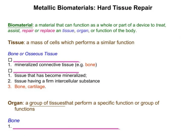
Hard tissue formation and destruction • Hard tissue associated with the functioning tooth: cementum, dentin, and enamel • Specialized connective tissues (except enamel) • Collagen (esp. type I) plays a role in determining their structure. • Hard tissue formation: cells - production of organic matrix -capable of accepting mineral + the activity of the enzyme alkaline phosphatase + a good blood supply prerequisites.
Cells • functions of synthesis and secretion • abundant mitochondria, much rER, and Golgi complex Organic matrix • type I collagen + proteoglycans + phosphoproteins + phospholipids • enamel: enamel proteins: amelogenin + enamelin • capable of accepting mineral in the form of hydroxyapatite crystals
Mineral • biological apatite: calcium hydroxyapatite [Ca10(PO4)6(OH)2] • calcium + phosphate + hydroxyl ions - ionic lattice relationships - permit considerable variation in composition through substitution, exchange, and adsorption of ions • magnesium and sodium can substitute in the calcium position • fluoride and chloride can substitute in the hydroxyl position • carbonate can substitute in both the hydroxyl and phosphate positions • fluoride substitution - decrease the solubility of the crystallites • carbonate substitution - increase the solubility of the crystallites
Mineralization • local increase in the concentration of inorganic ions - supersaturated tissue fluid of calcium and phosphate ions - spontaneous precipitation of a calcium phosphate product - formation of ionic clusters and crystallites (homogeneous nucleation) • the presence of a nucleating substance but not locally increased ionic concentration - crystal formation (heterogeneous mineralization)
factors inhibit mineralization • tissue fluid contains other macromolecules that could inhibit crystal formation • the initial cluster of ions needed to form a lattice structure is not enough or unstable • insufficient energy for overcoming crystallization (inhibited by inhibitors of mineralization)
Bone • Three functions • Mechanical: support and site of muscle attachment for locomotion • Protective: for vital organs and bone marrow • Metabolic: reserve of ions for the entire organism, especially calcium and phosphate
Bone physiology • Non cellular elements: • 33% organic: • 28%collagen fiber type I, • 5% ground substance : glycoproteins, proteoglycans. These highly anionic complexes have a high ion-binding capacity and are thought to play an important part in the calcification process and the fixation of hydroxyapatite crystals to the collagen fibers. • 67% inorganic:hydroxyapatite.
Bone structure • Lamellar structure: this organization of fibers allows the highest density of collagen per unit volume of tissue. These lamellae can be parallel to each other if deposited along a flat surface (trabecular bone and periostrum) or concentric if deposited on the surface of a channel centered on a blood vessel (Haversian system) • Woven bone: when bone is formed very rapidly (during histogenesis, fracture healing, tumor, or some metabolic bone disease). They are found in more or less randomly oriented bundles.
Cellular elements • Osteocytes • Osteblasts • Osteoclasts • Other cell types
Osteocytes • They are found embeded deep within the bone in small osteocytic lacunae (25000/mm3 of bone). • They were originally bone-forming cells (osteoblasts) that have been trapped into their own production of bone matrix, which later became calcified. • These cells have numerous and long cell processes rich in microfilaments that are in contact with cell processes from other osteocytes (gap junctions), or with processes from the cells lining the bone surface (osteoblasts or flat lining cell in the endosteum or periosteum) • These processes are organized during the formation of the matrix and before its calcification.
Osteblasts • The osteoblast is the bone lining cell responsible for the production of the matrix constituents (collagen and ground substance). • It originates from a local mesenchymal stem cell (bone marrow stromal stem cell or connective tissue mesenchymal stem cell) • These precursors upon the right stimulation, undergo proliferation and differentiate into preosteoblasts and then mature osteoblasts.
Osteblasts • Osteoblasts never appear or function individually but are always found in clusters of cuboidal cells along the bone surface (~100-400 cells per bone froming site). • They are always found lining a layer of bone matrix that are producing and is not yet calcified (osteoid tissue, its maturation period ~10 days)
At the ultrastructural level • the osteoblastis characterized by • The presence of an extremely well-developed rough endoplasmic reticulum and a dense granular content • The presence of a large circular Golgi complex comprising multiple Golgi stacks
Osteblasts • Cytoplasmic processes on the secreting side of the cell extend deep into the osteoid matrix and are in contact with the osteocyte processes in their canaliculi. • Junctional complexs (gap junctions) are often found between the osteoblasts. • The plasma membrane of the osteoblast is characteristically rich in alkaline phosphatase (used as an index of bone formation) and been shown to have rceptors for parathyroid hormone, but not for calcitonin.
Osteblasts • OBs arise from osteoprogenitor cells of mesenchymal origin and are the source of the terminally differentiated osteocyte • OBs have the ability of synthesize type I collagen and regulates its mineralization into a specific crystalline form • OBs are autocrine regulatory cells. They can synthesize and deposit growth factors in bone matrix which when released by bone resorption processes, restimulate further OB activity • OBs mediate the systemic signals for the recruitment and activity of OCs.
Osteblasts • Osteoblasts also express receptors for estrogens and Vit D3 in their nuclei. • Toward the end of the secreting period the osteoblasts will become either a flat lining cell or an osteocyte
Bone formation • The formation of bone by mesenchymal cells can occur by one of 2 routes • A direct development of bone from mesenchymal cells, as in the calvarium, called intramembranous ossification • An intervening cartilage model precedes the formation, ie the proliferation of mesenchymal cells is followed by their differentiation into chondrocytes which become hypertrophied and calcified. The calcified cartilage is replaced by bone which is then remodelled (endochondral ossification)
Bone formation • Synthesis and intracellular processing of type I collagen • Secretion and extracellular processing of the collagen • The formation of microfibrils, fibrils, and ultimately fibers from the collagen • Maturation of collagen matrix with subsequent nucleation and growth of HA crystals • OCs also plays an important role in development and growth of bone by releasing polypeptide growth factors from the extracellular mineralized matrix (bone-derived growth factors, BDGFs)
Fig. 1-81 illustrates an osteon with osteocytes (OC) residing in osteocyte lacunae in the lamellar bone. The osteocytes connect via canaliculi (can) which contain cytoplasmatic projections of the osteocytes. A Haversian canal (HC) is seen in the middle of the osteon. Fig. 1-82 illustrates an area of the alveolar bone in which bone formation occurs. The osteoblasts (arrows), the bone-forming cells, are producing bone matrix (osteoid) consisting of collagen fi bers, glycoproteins, and proteoglycans. The bone matrix or the osteoid undergoes mineralization by the deposition of minerals such as calcium and phosphate, which are subsequently transformed into hydroxyapatite. Fig. 1-83 The drawing illustrates how osteocytes, present in the mineralized bone, communicate with osteoblasts on the bone surface through canaliculi.
The cellular events involved in bone formation • Cessation of continued OCs activity and disappearance of multinucleated OCs from resorptive site • Attraction of OB precursors to the site • Proliferation of OB precursors • Differentiation of these precursors to form mature collagen-secreting OBs • Formation of mineralized bone matrix • Cessation of OB activity
Cessation of OCs activity • Some OCs undergo a process of programmed cell death or apoptosis at remodeling sites, and then presumably removed by scavenger cells • The constituents of mineral phase of the bone matrix could be involved in inhibition of continued OCs activity: • Free ionized calcium as well as phosphate • TGF-b which is an inhibitor of OC activity and is a powerful enhancer of OC apoptosis may be produced by OCs, by OBs in the neighborhood of OCs, or be released from the bone matrix itself.
Chemotaxis • Chemotactic signals could include constituents of the bone matrix such as fragments of collagen or osteocalcin, or growth factors produced in active from at the resorption site such as TGF-b and PDGF • include the attraction of OB precursors to the site of resorption by a process of chemotaxis, their stimulation is followed by differentiation to mature cells which are capable of synthesizing bone protein.
Proliferation • Bone cell mitogens include insulin-like growth factor (IGF-I, II), platelet-derived growth factor (PDGF), and the heparin-binding fibroblast growth factors (FGFs). • transforming growth factor-b (TGF-b) is a powerful bone cell mitogen • These factors could stimulate to OB precursor proliferation.
Differentiation • The factors involved in stimulation of differentiation of OB to form mature OBs. • bone morphorgenic proteins (BMPs) are powerful differentiation agents • The only other agents which are known to have similar effects are fluoride, 1,25 vitD, and retinoic acid • OBs synthesis and secrete the extracellular organic matrix of bone, including type-1 collagen, osteocalcin, osteopontin, alkaline phosphatase, proteoglycans as well as the growth regulatory factors.
Mineralized bone matrix • initial mineralization: involving matrix vesicle • small, membrane-bound vesicle - providing a microenvironment for initial mineralization • containing alkaline phosphatase, pyrophosphatase, Ca-ATP-ase, metalloproteinase, proteoglycans, and anionic phospholipids - bind to calcium and inorganic phosphate - form calcium-inorganic phosphate phospholipid complexes (must maintain) • deposition of apatite crystallites in relation to the collagen fibrils and noncollagenous proteins
Alkaline phosphatase • may or may not associated with matrix vesicles • function: hydrolyzing phosphate ions from organic radicals at an alkaline pH • nonspecific; may have more than one distinct function in mineralization • breaking down pyrophosphate -removing its inhibition of hydroxyapatite crystal growth - permitting crystal growth to proceed
Regulatory factors involved in normal bone formation • TGF-b is one of the most abundant of the growth regulatory factors in bone matrix. • TGF-b exerts its major effect on bone formation by stimulating proliferation of OB precursors and increasing the pool of committed OBs, and it also increases OB chemotaxis. • BMPs are growth regulatory factors in extended TGF-b family, and in particular BMPs 2,3, and 4 are expressed by OBs as they differentiate. • BMP2 and BMP4 seem to have effects on bone cell differentiation
heparin-binding fibroblast growth factors (FGFs) are powerful bone forming factors. Both acidic and basic FGFs stimulate bone formation in vivo which are probably mediated predominately by stimulation and proliferation of OB precursors. • PDGF is expressed by bone cells and there are also receptors for PDGF on bone cells
Cascade theory of bone formation • In the early phases, this probably involves a number of chemotactic and proliferative factors such as PDGF, TGF-b and IGF. • As these cells multiply, they express mitogenic growth factors such as TGF-b, PDGF and the FGFs. • As the cells starts to differentiate, they express a number of genes involved in the bone formation process such as osteoclacin, alkaline phosphatase, and type I collagen. • They also express the BMPs, which are autostimulatory. As a consequence, expression of BMPs may be responsible for the final stages of bone cell differentiation at the formation of normal mineralized bone.
Osteoclasts and bone resorption • The osteoclast is the bone lining cell responsible for bone resorption • It is a giant multinucleated cell (4-20 nuclei) usually found in contact with a calcified bone surface and with a lacuna (Howship’s lacunae), which is the result of its own resorptive activity. • Possible 4 or 5 osteoclasts in the same resorptive site, but usually only 1 or 2 per site.
The cytoplasm is “foamy” with many vacuoles • The contact zone with bone is characterized by the presence of a ruffled border and dense patches on each side of it known as the sealing zone.
At the ultrastructural level • The abundance of Golgi complexs characterically disposed around each nuclei, mitochondria, and transport vesicles loaded with lysosomal enzymes. • The attachment of the cell to the matrix is performed via integrin receptors, binding to specific sequences in matrix proteins.
Mononuclear-phagocyte lineage are the most likely candidates to differentiate into osteoclasts. May occur at the promonocyte stage, but monocytes and macrophages, already commited to their own lineage, might still be able to form osteoclasts under the right circumstances. Origin of the osteoclast
Resorptive events • attachment of osteoclasts to the mineralized surface of bone • creation of a sealed acidic environment through action of the proton pump, which demineralizes bone and exposes the organic matrix • degradation of this exposed organic matrix to its constituent amino acids by the action of released enzymes such as acid phosphatase and cathepsin B • uptake of mineral ions and amino acids by the cell
Mechanisms of bone resorption • Lysosomal enzymes: in the endopalsmic reticulum, Golgi, and many transport vesicle; these lysosomal enzymes are secreted, via the ruffled border, into the extracellular bone resorbing compartment. • Nonlysosomal enzymes: metalloproteinase such as collagenase, stromelysin and elastase. All these proteinases would not retain much activity in the low pH of the ruffle border.
Mechanisms of bone resorption • Proton pumps: the presence of an electrogenic proton pump ATPase. • The protons are provided to the pumps by the enzyme carbonic anhydrase, highly concentrated in the cytosol of this cell, and ATP and CO2 are provided by mitochondria. • The basolateral membrane activity exchanges bicarbonate for chloride (HCO3-/Cl-), thereby avoiding and alkalinization of cytosol. • The basolateral sodium pumps might be involved is secondary active transport of calcium and/or protons in association with Na+, Ca++ exchanger and/ or Na+, H+ antiport
Extracellular bone resorbing compartment • A low pH: dissolves the crystals, exposing the matrix • Lysosomal enzymes: now at optimal pH, degrade the matrix components. • Substrate: the residues from this extracellular digestion are either internalized, or transported across the cell (transcytosis) and releasesed at the basolateral domain, or else released during periods of relapse of the sealing zone, possibly induced by a calcium sensor in response to the rise via extracellular calcium in the bone resorbing compartment.
Bone remodeling • A balance between synthesis and breakdown is now widely called “coupling” of bone resorption and formation. • Resorption involves three steps: • The bone resorbing agent, PTH, induces a change of shape of the osteoblast, thus facilitating the access of osteoclasts to the bone surface. • The osteoblasts systhesis the collagenase and plasiminogen activator which digests the osteoid, exposing the mineralized matrix, which may be chemotactic to the OC. • The osteoblasts release the short-range soluble activator to the OC.
Remodeling of the trabecular bone starts with resorption of the bone surface by osteoclasts (OCL) as seen in Fig. 1-90a. After a short period, osteoblasts (OB) start depositing new bone (Fig. 1- 90b) and fi nally a new bone multicellular unit is formed, clearly delineated by a reversal line (arrows) as seen in Fig. 1-90c.
The osteoblast-osteoclast relationship • Osteoblast contains the receptors for the major bone resorbing agents such as, PTH, the eicosanoids (PGE2, PGI2, LTB4), 1,25 dihydroxyvitamin D3, and cytokines (IL-1, TNF), and transmit resorptive signal to the osteoclast. • OBs secrete a short-range souble activator (lipoxygenase metabolites of arachidonic acid) for OCs • OBs appear to play a pivotal role in the regulation of bone resorption.
Biology of bone healing (granulation tissue)
Initial injury - Caused extravasation and cell signaling • Proteolytic degradation of ECM produces chemotactic remnants, luring monocytes and macrophages to wound bed • Activated macrophages release FGF, stimulating endothelial cells to express plasminogen activator and procollagenase. • Growth factors released from the a granules of degranulating platelets are beacons for PMNs, lymphocytes, monocytes, and macrophages.
Proliferation • Granulation tissue develops consisting of new blood vessels, collagen isotypes and cells. • At least 18 isotypes of collagens: type I is associated with bone, type II with cartilage, types III and V with granulation tissue, types IV and VI with the endothelial matrix, and type X with hypertrophic cartilage. • Collagenous substratum which is a key instructional substratum of the repairing wound functions as a provisional solid-state matrix for cellular attachment and seletive binding of growth factors. • The functional role of a callus is to stabilize the bone fragments
Proliferation • At the injury locus, the lure for monocytes that will convert into osteoclasts appears to be fragments of fibronectin and degradation products from extracellular matrix. • Macrophages at the wound site express FGF and VEGF, prompting neoangiogenesis.
Remodeling • The processes associated with hemostatic remodeling are known as activation-resorption-formation.The processes will take between 3 to 6 months • Osteoblasts are activated by signaling factors (PTH). • Osteoclasts home in to the osteoblast vacant zone, attach, resorb an in response to an as yet unidentified signal, cease resorbing and abandon their attachment. • Osteoclastic resorptive pits become repopulated by a contigent of osteoblasts that express osteoid, which calcifies, retsoring bone.
The mediators of bone remodeling • Parathyroid hormone • Parathyroid hormone related peptide (PTHrP) • Vitamin D metabolites • The cytokines • Eicosanoids • Growth factors • Bacterial products • Mechanical stress