Download
1 / 23
340 likes | 2.24k Views
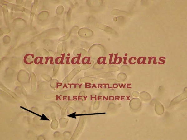
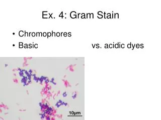
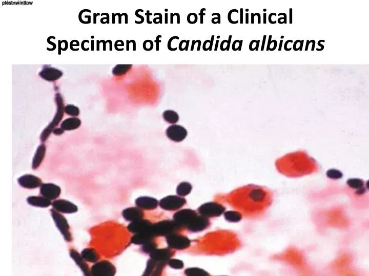
Gram Stain of a Clinical Specimen of Candida albicans. Growth on Sabouraud's Dextrose Media. تتحدث عن الصورة في الشريحه التاليه. Gram Stain Gram + stain of cocci arranged in culster. Staphylococcus. Stained in Pus. Staphylococci. Gram-Positive Streptococcus.

E N D
تتحدث عن الصورة في الشريحه التاليه • Gram Stain • Gram + stain of cocci arranged in culster
Stained in Pus Staphylococci
Sputum stained with gram positive microorganism pus
Staphylococcus Streptococcus
من القاموس A bacterium that causes the formation of pus or of fatal septicemias Streptococcus pyogenes = Group A Strep Carried by many people in throat or on skin often no symptoms Cause of • strep throat • Impetigo اصابه الجلد • Necrotizing موت الانسجه fasciitis
Streptococcus pyogenes. Left. Gram stain of Streptococcus pyogenes in a clinical specimen. Right. Colonies of Streptococcus pyogenes on blood agar exhibiting beta (clear) hemolysis
احفظ الانواع المسببه مهمه Alpha-hemolysis • Alpha-hemolytic Streptococcus species "Viridans group" streptococci, including species such as the Streptococcus mutans, mitis, and salivariusgroups display alpha hemolysis.
Alpha-hemolytic Streptococcus species Viridans group" streptococci, including species such as the Streptococcus mutans, mitis, and salivariusgroups display alpha hemolysis. (Also see Figures 15,16 and 23-25).
Demonstrations and practial • Bacitracinاسم مضادsensitivity الدائره هي العلامه الفارقه • Principle: • Bacitracin test is used for presumptive للترجيح identification of group A • To distinguish between S. pyogenes (susceptible حساسto Bacitracin) & non group A such as S. agalactiae (Resistant to B) • Bacitracin will inhibit the growth of gp A Strep. pyogenes giving zone of inhibition around the disk • Procedure: • Inoculate يلقح BAP الاجار ( الميديا اللي فيها المضاد )with heavy suspension of tested organism • Bacitracin disk (0.04 U) is applied to inoculated BAP العينه الملقحه • After incubation فتره الحضانه, any zone of inhibition around the disk is considered as susceptible حساس من هذا المضاد يعني ( قروب أي ) Group A
اختبار نفس فكره الاختبار اللي راح بس البكتيريا الحساسه هنا هي : s.pneumonia Optochin resistant S. viridans Optochin resistant S. viridans Optochin susceptible S. pneumoniae
Lowenstein-Jensen medium Lowenstein-Jensen medium Mycobacterium tuberculosis Name of its medium Mycobacterium tuberculosis
Aspergillusniger Culture of Aspergillusniger. Conidial head of A. niger
Streptococcus pneumoniae - optochin sensitivity test alpha hemolysis (green zone surrounding colonies). Note the zone of inhibition around a filter paper disc impregnated وضع عليهwith optochin. Viridans streptococci are not inhibited by optochin.
Haemophilus Species Direct smear لطاخهof H. influenzae in CSF in a case of meningitis. Note the intracellular and extracellularpleomorphicمتعدد الاشكالgram-negative bacilli.