Download
1 / 1
10 likes | 16 Views
This study examines the genetic resistance to Soybean Mosaic Virus in Jindou 1 Soybean through the identification of molecular markers associated with resistance alleles.

E N D
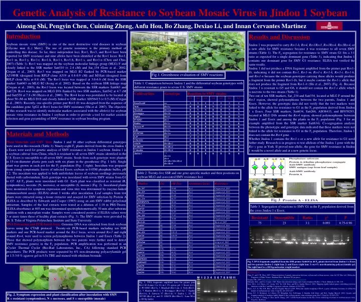
Genetic Analysis of Resistance to Soybean Mosaic Virus in Jindou 1 Soybean Ainong Shi, Pengyin Chen, Cuiming Zheng, Anfu Hou, Bo Zhang, Dexiao Li, and Innan Cervantes Martinez Introduction Soybean mosaic virus (SMV) is one of the most destructive viral diseases in soybean [Glycine max (L.) Merr.]. The use of genetic resistance is the primary method of controlling this disease. So far, three independent loci, Rsv1, Rsv3, and Rsv4 have been reported for SMV resistance and nine alleles have been identified at the Rsv1 locus: Rsv1, Rsv1–m, Rsv1-y, Rsv1-r, Rsv1-k, Rsv1-t, Rsv1-h, Rsv1-s, and Rsv1-n (Chen and Chio, 2007) (Table 1). Rsv1 was mapped on the soybean molecular linkage group (MLG) F and closely linked to the SSR marker Satt510 (< 2.4 cM) and Sat_154 (Gore et al., 2002; Cregan et al., 2003). Rsv3 was mapped on MLG B2 flanked by PCR-based marker A519F/R (designed from RFLP clone A519 at 0.8-0.9 cM) and M3Satt (designed from RFLP clone M3a at 0.8 cM). The Rsv3 locus was mapped at 3.0-6.0 cM from the SSR marker Satt063 on MLG B2 (Jeong et al. 2002). Based on the soybean SSR linkage map (Cregan et al., 2003), the Rsv3 locus was located between the SSR markers Satt063 and Satt726. Rsv4 was mapped on MLG D1b flanked by two SSR markers, Satt542 at 4.7 cM and Satt558 at 7.8 cM (Hayes et al., 2000). The Rsv4 locus was postulated to be located at about 50 cM on MLG D1b and closely linked to SSR marker AI856415 (< 0.5 cM) (Cregan et al., 2003). Recently, one specific primer pair Rsv1-f/r was designed from the sequence of the candidate gene 3gG2 at Rsv1 locus for SMV resistance (Shi et al. 2007). The objective of this research was to identify molecular markers associated with the allele(s) for soybean mosaic virus resistance in Jindou 1 soybean in order to provide a tool for marker assisted selection and gene pyramiding of SMV resistance in soybean breeding program. Results and Discussion Jindou 1 was proposed to carry Rsv1-h, Rsv4, Rsv1Rsv3, Rsv1Rsv4, Rsv3Rsv4, or a new allele for SMV resistance because it was resistance to all seven SMV strains (Table 1). The F2 segregation for resistance to SMV strain G1 fits a 3:1 ratio as expected for a single dominant gene (Table 3), indicating that Jindou 1 contains one dominant gene for SMV G1 resistance. ELISA test verified the same results. Jindou 1 did not produce a DNA fragment amplified from the primer pair Rsv1-f/r, indicating it did not contain Rsv1, Rsv1–m, Rsv1-r, Rsv1-k, Rsv1-t, Rsv1-h, and Rsv1-n because the soybean genotypes carrying these alleles would produce a fragment from the primer Rsv1-f/r, but it maybe contain the Rsv1-y allele that does not produce the fragment from Rsv1-/r (Shi et al., 2007) (Fig. 4). Because Jindou 1 is resistant to G5 and G6, it should not contain the Rsv1-s allele which is necrotic to the two strains (Table 1). Two out of ten SSR markers, Sat_234 and Satt334, located at MLG F around the Rsv1 region, showed polymorphisms between the two parents, Jindou 1 and Essex. However, the genotypic data did not verify that the two markers were linked to the allele for resistance to G1 in the F2 population derived from Jindou 1 x Essex. Four SSR markers, Satt634, Satt266, AI856415, and BF070293, located at MLG D1b around the Rsv4 region, showed polymorphisms between Jindou 1 and Essex and among the plants in the F2 population (Fig. 5 for an example amplified from the SSR marker Satt634). Co-segregation analysis between the phenotypic and genotypic data indicated that these markers were not linked to the allele for resistance to G1 in the F2 population. Therefore, Jindou 1 does not contain the Rsv4 gene. Whether Jindou 1 contains the Rsv1-y or a new allele for resistance to G1 need futher study. Research is in progress to test allelism of the Jindou 1 gene with the Rsv-y gene in York. If proved non-allelic, the gene for SMV resistance in Jindou 1 would be a novel allele and at a new genetic locus. Fig.1. Greenhouse evaluation of SMV reactions Table 1. Comparison between Jindou 1 and the differential soybean genotypes with different resistance genes to seven U.S. SMV strains Materials and Methods Plant Materials and SMV TestsJindou 1 and 16 other soybean differential genotypes were used in this research (Table 1). Ninety-eight F2 plants derived from the cross Jindou 1 x Essex were used for genetic analysis of SMV resistance in Jindou 1 soybean. Jindou 1 is a soybean cultivar from China, which is resistant to all seven SMV strains identified in the U.S. Essex is susceptible to all seven SMV strains. Seeds from each genotype were planted in 15-cm-diameter plastic pots each with six plants in the greenhouse (Fig. 1 left). Single plant was grown in each pot for the F2 population (Fig. 1 right). Inoculum was prepared from young symptomatic leaves of infected Essex soybean in 0.05M phosphate buffer, pH 7.2. The inoculum was applied to both unifoliolate leaves of soybean seedlings previously dusted with carborundum. Each genotype was inoculated with seven SMV strains from G1 to G7. All F2 plants were inoculated with G1. Each plant was classified as resistant (R, symptomless), necrotic (N, necrosis), or susceptible (S, mosaic) (Fig. 2). Inoculated plants were monitored for symptom expression and virus titer was determined by enzyme-linked immunosorbent assays (ELISA) about 3 weeks after inoculation. Leaf samples from test plants were extracted using a tissue extractor and assayed for SMV infection by Protein A ELISA as described by Edwards and Cooper (1985) using an anti-SMV rabbit polyclonal antiserum. Samples of the leaf extracts were tested at a dilution of 1:10 in PBS-Tween. ELISA absorbance at 405 nm was determined spectrophotometrically 30 min after substrate addition with a microplate reader. Samples were considered positive if ELISA values were 3 or more times those of healthy plant extracts (Fig. 3). The SMV strains were provided by Dr. S. Tolin of Virginia Polytechnic Institute and State University. DNA Extraction and PCR-based AssayGenomic DNA was extracted from fresh soybean leaves using the CTAB protocol. Twenty-six PCR-based markers including ten SSR markers and one PCR-based marker around the Rsv1 locus, seven around Rsv3 and eight around Rsv4, were used to screen polymorphisms between Jindou 1 and Essex (Table 2). Those that showed polymorphism between the two parents were further used to detect SMV resistance gene(s) in the F2 population. PCR amplification was performed in an iCycler Thermal Cycler (Bio-Rad Laboratories, Inc., CA) following standard PCR procedures. The PCR products were separated by 6% non-denaturing polyacrylamide gel or 1.5-3.0 % agarose gel in 0.5x TBE and stained with ethidium bromide. Table 2. Twenty-five SSR and one gene-specific marker and their positions on soybean MLG and associated SMV resistance loci Fig. 3 Table 3. Segregation of reactions to SMV G1 in the F2 population derived from the cross Jindou 1 x Essex. Fig. 5. DNA fragments amplified from the SSR primer Satt634 in 46 F2 plants derived from Jindou 1 x Essex and two parents Jindou 1 (right lane 2) and Essex (right lane 3) on 6% non-denaturing polyacrylamide gel. The right lane1 is a 100 bp molecular-weight marker R S References Chen, P., and C.W. Choi. 2007. Characterization of genetic interaction between soybean and soybean mosaic virus. In G.P. Rao (ed.) Molecular Diagnosis of Plant Viruses. Studium Press, Houston TX (in press). Cregan, P.B. 2003. (http://bldg6.arsusda.gov/~pooley/soy/cregan/cregan.htm). Edwards, M.L., and J.L. Cooper. 1985. Plant virus detection using a new form of indirect ELISA. J. Virol. Methods 11:309–319. Gore, M.A., A.J. Hayes, S.C. Jeong, Y.G. Yu, G.R. Buss, and M.A. Saghai Maroof. 2002. Mapping tightly linked genes controlling potyvirus infection at the Rsv1 and Rpv1 region in soybean. Genome 45:592-599. Hayes, A.J., G. Ma, G.R. Buss, and M.A.Saghai Maroof. 2000. Molecular marker mapping of Rsv4, a gene conferring resistance to all known strains of soybean mosaic virus. Crop Sci. 40:1434-1437. Jeong, S.C., S. Kristipati, A.J. Hayes. P.J. Maughan, S.L. Noffsinger, I. Gunduz, G.R. Buss, and M.A. Saghai Maroof. 2002. Genetic and sequence analysis of markers tightly linked to the soybean mosaic virus resistance gene, Rsv3. Crop Sci. 42:265-270. Shi, A., P. Chen, C. Zheng, A. Hou, and B. Zhang. 2007. A PCR-based marker for the Rsv1 locus conferring resistance to soybean mosaic virus. Crop Sci. (In Press). M 1 2 3 4 5 6 7 8 9 10 11 N Fig. 4. DNA fragments amplified from the primer pair Rsv1-f/r (Lanes 1 to 11) on 1.3% agrose gel: 1. Essex (rsv), 2. PI 96983 (Rsv1), 3. Marshall (Rsv1-m), 4. York (Rsv1-y), 5. Raiden (Rsv1-r), 6. Kwanggyo (Rsv1-k), 7. Ogden (Rsv1-t), 8. Suweon 97 (Rsv1-h), 9. Jindou 1, 10. PI 507389 (Rsv1-n), and 11. OX670 (Rsv1Rsv3) . Lane M is a 100 bp ladder Fig. 2. Symptom expression and plant classification after inoculation with SMV R = resistant (symptomless), N = necroses, and S = susceptible (mosaic)