Download
1 / 34
490 likes | 898 Views
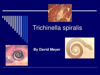
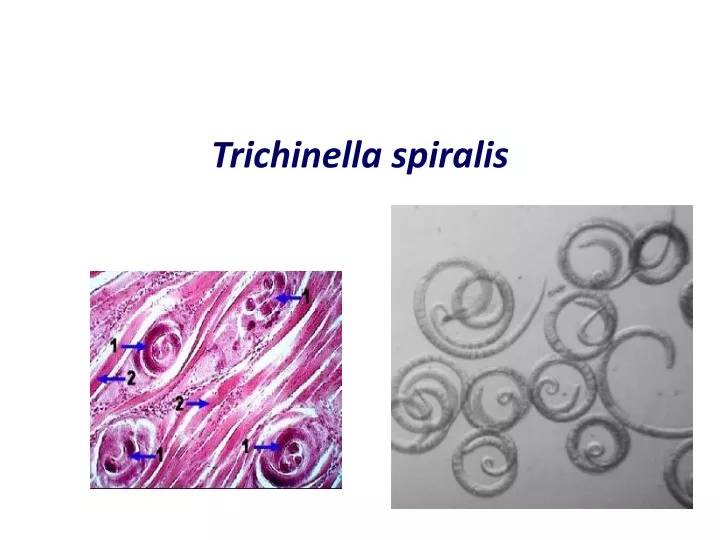
Trichinella spiralis. SALIENT FEATURES. COMMON NAME: Trichina worm - The Pork worm Trichinella spiralis means spira , how this coils up in its host DISEASE : Trichinosis It is a zoonotic disease HABITAT : Adult worms -Mucosa of small intestinal

E N D
SALIENT FEATURES • COMMON NAME: Trichina worm- The Pork worm • Trichinellaspiralismeans spira, how this coils up in its host • DISEASE: Trichinosis • It is a zoonotic disease • HABITAT: Adult worms-Mucosa of small intestinal Encysted larvae- Striated muscles of hosts • The same animal acts as definitive and intermediated host • Is most common in Europe, North America, and Asia • INFECTIVE STAGE: Contaminated meat (muscle) containing encysted larvae (pig) • DIAGNOSTIC STAGE: Larvae encysted in muscle (human) • Can be fatal if large numbers of cysts form in the heart muscle.
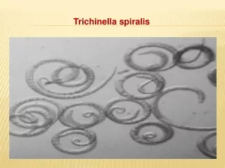
VIVIPAROUS Expel active larvae
Epidemiology • The disease occurs among pigs, rats & humans • Rats and pigs feeding on garbage that includes infected offal • Dead or dying infected rats are themselves eaten by pigs • Raw or poorly cooked pork (sausage) harboring infective larvae then become the vehicle for human infections • Trichinellosis is a cosmopolitan disease that occurs most commonly in Europe and the US
PATHOGENESIS: Penetration of the adult females into mucosa The first symptoms appear between 1- 2 days after ingestion The worms migrate in the intestinal epithelium Inflammation of duodenal and jejunal mucosa This causes: • INFLAMMATION • NAUSEA • VOMITING • SWEATING • DIARRHEA
The migrating larvae Ten days after infection the larvae will penetrate the muscle fibers & other parts of the body. • Muscular pain • Difficulty breathing • Per orbital edema and conjunctivitis • Heart (Myocarditis) • Lungs (Pneumonitis) • Brain (Encephalitis) • Can be fatal if large numbers of cysts form in the heart muscle. • Heart failure or respiratory or kidney malfunction
DIAGNOSIS : • Muscle biopsy at the encystment stage • Blood test for eosinophilia • Increased levels of creatinephosphokinase(CPK) • Serology test Immunoassays, such as ELISA • At the diarrheal stage, adult and larvae may be found in faeces
TREATMENT • THIABENDAZOLE • MEBENDAZOLE • CORTICOSTEROIDS
Trichuris trichiura Whip worm
Trichuristrichiura • Common name: whip worm • Disease:trichuriasis, whip worm infection • Final host: human, dogs, pig, monkey • Habitat: large intestinal ( cecum, appendix, rectum) • Geographical distribution: Cosmopolitan with poor sanitation. • Children are more likely to be infected than adults because they are more likely to have have close physical contact with contaminated soil • Infective stage: infective larva in egg • Transmission occurs through ingestion of eggs, usually on contaminated vegetables or soil. • Diagnostic stage: Egg barrel shape with polar plugs
Morphology: Adult female worm: The anterior two-thirds of the body being very thin (looks like a whip) and the remaining posterior end is thick and linear. Size: 3.5-5cm in length Adult Male worm: smaller than the female, 3.0-3.5cm. The posterior end is curved and has a single spicule enveloped with sheath.
The anterior end two-thirds of the body being very thin (looks like a whip). • Adult worm penetrates into and embed its whip-like anterior portion in the intestinal mucosa, By small spear Adult male Adult female • Longer than the male.- posterior end is thick • and linear. • Shorter than the female. posterior end curved and • has a single spicule • enveloped with sheath. • .
posterior end curved and has a single spicule enveloped with sheath
Eggs: Shape: barrel–shaped Size: 50-55 x 25-30μm Shell: thick egg shell with 2 polar plugs Color: Yellow-brown Content: immature egg cells 3000-10000 eggs daily daily output
Life cycle: • Eggs pass out immature • Embryo develops inside the egg (that takes about 3weeks at 25C) • Mature eggs swallowed 1st stage larvae hatch in small intestine and penetrate villi • Then migrate to large intestine and attach to mucosa with the thin anterior end • After 2-4 month females mature and lay eggs.
Pathology: • Light infection with Trichiuris are asymptomatic • Heavier infections are characterized by 1- diarrhea, 2- anorexia, 3- nausea 4- abdominal pain 5- anemia may be the result of hemorrhaging when the worms mucosal damage))penetrate the intestinal wall • Rectal prolapse. Children’s infection can cause rectal prolapse, The reason is the cecum is damaged by the worm, the cecum can be pushed out from the anus.
Laboratory diagnosis 1- Eggs or worm in feces. Eggs are oval, barrel shaped, 2- Eosinophilia may occur. 3- In heavy infection proctoscopy or sigmoidoscopy,can show the worms attached to the mucosa. 4- Visual detection of adult worms on prolapsed rectum.