Download
1 / 16
160 likes | 255 Views
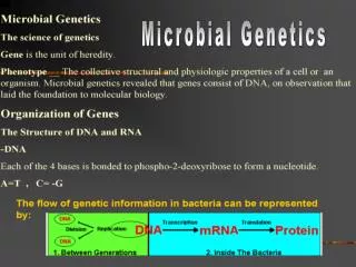
Microbial Genetics Part 2. Genetic Mutation. A genetic mutation is a change in the original DNA nucleotide sequence. It can consist of a change in one or more base pairs; A deletion of one or more base pairs; An addition of one or more base pairs.

E N D
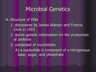
Genetic Mutation • A genetic mutation is a change in the original DNA nucleotide sequence. • It can consist of a change in one or more base pairs; • A deletion of one or more base pairs; • An addition of one or more base pairs. • Some mutations harmful, some beneficial, some neutral. • A point mutation is one in which a single base pair is changed, added, or deleted. • It can cause a change in the codon sequence, thereby changing the codons to code for different amino acids. It could even change the codon to a stop codon in middle of the protein resulting in a shortened and probably non-funtional protein. (This type of mutation is called a nonsense mutation.)
A point mutation can cause a change in overall shape and function of the final protein. An example of a point mutation is sickle cell anemia. The point mutation changes the shape of the red blood cell so that it cannot function correctly. Ironically, it is this change of shape that often protects Africans from contracting Malaria which is so common on that continent. It is thought that this particular mutation developed as a result of the high exposure to Malaria as a natural defense against the disease. (In other words, an example of evolution.) • A frameshift mutation is one in which a one or more base pairs are added or deleted. • When 1 base pair is added into the DNA sequence the effects could be huge! If the base pair were added into the 1st position of the codon then the whole sequence of the following codons are changed. Potentially every single amino acid would be different from what the original code specified. • It is called a frameshift mutation because it cause a literal shift in the reading frame of each subsequent codon. • Another type of mutation is a silent mutation. A silent mutation changes a base pair but the change in the base pair does not alter the amino acid coded for by the codon. • Take a look at Fig. 9.14 on page 264. Look for the amino acid, Glycine. You’ll notice that Glycine has 4 different codons that code for it. So, if a mutation in the DNA template changed GGU to GGC no harm has been done because the amino acid remains the same.
Of course any mutation that occurs can always mutate back to the original sequence. This type of mutation is called a back-mutation. • Mutagens are agents in the environment that directly or indirectly cause mutation. • UV light and benzpyrene (chemical in smoke and soot) cause frameshift mutations. • Cells do have proofreading and repair enzymes to help take care of mutations. However, when the damage is widespread, the cells enzymes simply cannot take care of them all. If the damage is sever enough the cell will die.
Genetic Transfer and Recombination • Recombination is the exchange of homologous genes on a chromosome. • For example, when gametes are being formed the homologous chromosomes line up side by side. When they are in close proximity similar sequences of DNA can changes chromosomes. So if the chromosomes code for eye color but on one chromosome the gene was for brown eye color and on the other chromosome the gene was for green eye color, they could switch chromosome. • This DNA trading or switching is recombination. It is a very useful tool that we use in the laboratory too.
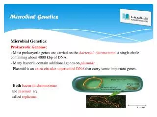
Often the question is asked, “How can bacteria become antibiotic resistant?” • One way bacteria gain antibiotic resistance by acquiring the resistance genes from other bacteria in the environment. • Bacteria acquire new genetic material in 4 ways that we know of currently. • Transformation • Conjugation • Transduction • Transposons
Transformation is when extracellular DNA is picked up by a bacterial cell. • After cell death, some bacteria are lysed (broken open) and release their cellular contents into the surrounding environment. • Some bacteria have the ability to take up that DNA and incorporate it into their chromosome or use it in some other way. • In order to take up extracellular DNA the cell has to be competent.
Competence means that the recipient cell is in a physiological state that will allow it to take up DNA. • Transformation occurs naturally among only a few organisms. (This is another important tool that is used in the laboratory.) • Some plasmids encode for genes that enhance the pathogenicity of a bacterium. • For example, some strains of E.coli contain a plasmid that codes for toxins. • Other plasmids code for antibiotic resistance.
Conjugation uses pili to attach to a neighboring bacterial cell and transfer DNA through it. • Conjugation requires cell to cell contact in order for the process to begin. In addiiton, both cells must be opposing mating types. • I’m sure that seems confusing since we already know that bacteria aren’t male or female. Not to worry, here is what that means. • One cell must have the plasmid (F factor plasmid) that codes for pili formation. This cell is called the F+ cell. (Remember that a plasmid is a small circular piece of DNA in addition to a chromosome.) • The other cell must not have theF factor plasmid in order for conjugation to occur. • See Fig.9.24 for a great diagram and explanation of this process.
Steps of conjugation • 1. The pilus of the F+ cell attaches to the F- cell. • 2. The F factor plasmid begins to replicate. • 3. The plasmid copy begins to move through the pilus from the F+ cell to the F- cell. • 4. The connection between the two cells is broken. • When the recipient cell receives a complete copy of the F factor plasmid, it is then called a F+ cell. • Sometimes the connection between the two cells is broken early and only part of the F factor plasmid is transferred. In those cases, the cell generally remains an F- cell because it cannot be a donor cell without the complete copy.
Occasionally the F factor plasmid will recombine into the recipient chromosome. When that happens the cell is called an Hfr. • Hfr= high frequency of recombination • It can still produce pili and attach to other recipient cells. • The difference is that when the donor begins to replicate the plasmid it goes on to replicate the chromosome as well. • Because the chromosome is so much larger than the F factor plasmid by itself, the connection between the two cells is usually broken before a complete copy of the plasmid and chromosome can be transferred. • The DNA that is transferred usually recombines into the recipient chromosome and any unused DNA is degraded.
Transduction • Transduction is a process that uses bacteriophages to transfer DNA. • Bacteriophages are viruses that infect bacteria. • See Fig. 9.26 for a great explanation of this process. (You can look at 9.27 for your information but you won’t be responsible for that.)
A brief overview of phage replication • 1. The phage attaches to the cell wall of the target/host bacteria. • 2. The phage injects or releases its DNA into the host bacteria. • 3. The phage takes over the host cell and uses it to replicate its own DNA and make its own proteins. • 4. While phage DNA is replicated, bacterial DNA is broken down to free up more nucleotides for more phage DNA. • 5. Phage body parts are made and assembled. • 6. The phage DNA is packaged into the assembled phage. • 7. The cell breaks open and releases the phages into the environment.
Steps in transduction • 1. Phage infects the host bacterial cell. • 2. During replication and assembly, a phage incorporates a portion of the host cell DNA into itself, instead of phage DNA. • 3. The cell breaks open and the phages are released. • 4. The phage that contains the bacterial DNA infects another bacterial cell. • 5. Instead of producing more phages, the bacterial DNA is incorporated into the host cell chromosome.
Transposons • Transposons were discovered by Barbara McClintok while studying color variations in Indian Corn. • She believed that the color variations were caused by “jumping genes” that would jump into or out of the middle of the chromosome. • Her theories were met with a great deal of criticism and weren’t accepted until almost 30 years later. • Transposons contain genes that enable the short segment of DNA to insert and remove itself from the host genome. • Transposons have been found to be more common than we originally believed. They are even found in the human genome. • Transposons are important because when they insert themselves into the host chromosome we never know where it will happen. They can turn genes on or turn them off just by where they insert themselves.
All of the ways in which DNA can be transferred from cell to cell are used in laboratories to manipulate bacteria. • Make sure that you understand them so that when we start the biotechnology chapter you will know what is going on. • The hw for microbial genetics is due Friday, Oct. 6, 2006 by midnight.