Download
1 / 49
510 likes | 717 Views
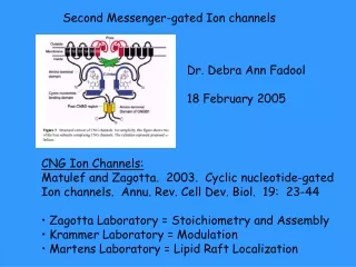
Second Messenger Systems. MECHANISM OF ACTION OF HYDROPHILIC MESSENGERS. Hydrophilic messengers cannot cross the cell membrane.

E N D
MECHANISM OF ACTION OF HYDROPHILIC MESSENGERS Hydrophilic messengers cannot cross the cell membrane. This restriction forces them into a 'classical' method of messenger action, namely the production of intracellular second messengers which are responsible for the effects of the various messengers.
Ligand binds to receptors located on the external surface of the plasma membrane. The resultant ligand·receptor complex is coupled to an enzyme located on the internal surface of the cell membrane and stimulates the conversion of some metabolite into a second messenger. In a large number of cases, the second messenger acts by stimulating or inhibiting protein phosphorylation/ dephosphorylation cascades. This second messenger then is responsible for the manifestations of all of the effects of the primary messenger inside the cell in question. Cascades have certain advantages to include signal divergence, convergence and amplification.
Hydrophilic messenger systems: G-Protein Systems As mentioned previously, hydrophilic messenger response systems require a transduction system to transfer the signal across the cell membrane to the cytoplasm. In many cases this transduction system involves a member of a family of proteins the guanyl nucleotide regulatory proteins or G-proteins. These proteins undergo a cyclic activation/inactivation which is controlled, to a certain extent, by ligand·receptor complexes.
G-proteins exist as heterotrimers with an , a and a subunit. The and subunits form a stable dimeric complex while the subunit contains a guanyl nucleotide binding site (for which the family is named). In the inactive state the nucleotide binding site is occupied by GDP.
Activation of the protein requires that the GDP be exchanged for GTP. This is accomplished by forming a trimeric G-protein complex which can associate with a receptor. When the receptor binds its ligand
The dissociation rate for GDP from the complex is increased and the nucleotide leaves the binding site.
Since GTP is present in cells at much higher concentrations and the affinity of the binding site for a triphosphate is higher, GTP binds to the complex. When GTP is bound to the nucleotide binding site of the subunit a number of changes/events occur.
The affinity of receptor for ligand decreases (so ligand tends to dissociate from receptor), the G-protein separates from the receptor and splits into an subunit and a dimer, and the -subunit becomes 'active'. In this state it can interact with various membrane proteins and alter their activities.
The -subunit also possesses an inherent GTPase activity which hydrolyzes the GTP to GDP and inactivates the G-protein. Thus the cycle has a built-in off switch.
The -subunit and the dimer each can exert biological effects. The type of G-protein and the action that it has is determined by the - subunit, and the sub-families are named based on their effects on the enzyme adenylyl cyclase which produces cAMP, the first second messenger discovered. Those G-proteins (-subunits) which stimulate adenylyl cyclase are referred to as Gs and those that inhibit are referred to as Gi while those that do neither are called Go.
Bacterial Toxins The discovery of the G-proteins and the elucidation of their mechanism of action was enhanced by the of various bacterial endo-toxins on the cycle. The two most important of these are cholera toxin and pertussis toxin. Both of these toxins ADP-ribosylate arginine residues in G-proteins and alter their activity. This reaction uses NAD+ as the ADP-ribose donor and releases nicotinamide.
Cholera toxin ADP-ribosylates the -subunits of Gs's and abolishes the GTPase activity of the subunit, creating a continually active Gs.
Pertussis toxin ADP-ribosylates the -subunits of Gi's and prevents the dissociation of GDP from the nucleotide binding site, creating a continually inactive Gi. Thus, the effect of either toxin is to stimulate the activity of the adenylyl cyclase system in target cells.
Adenylyl Cyclase System Cyclic AMP is generated by the enzyme adenylyl cyclase (AC), which in turn is subjected to both stimulatory and inhibitory input from ligand·receptor complexes via G-proteins (and other sources). As mentioned above, Gs has stimulatory effects on AC and Gi has inhibitory effects. The activation of the respective G-proteins is via two classes of receptors, one which acts on Gs and one which acts on Gi. This system uses cyclic AMP as the second messenger.
Cyclic AMP levels are also controlled by removal of the compound by the enzyme phosphodiesterase which converts the compound to AMP.
The effects of cAMP in cells are manifest through the action of particular protein kinases, protein kinase A's or cAMP-dependent protein kinases (CADPK's). These enzymes exist as hetero-tetrameres with two regulatory and two catalytic subunits. When bound together in a complex, the catalytic subunits are inactive.
The binding of cAMP to the regulatory subunit releases the catalytic subunits which are then free to phosphorylate serine and threonine residues in target proteins and alter the activity of the phosphorylated protein. The phosphorylated protein is converted back to its original state by the action of phosphoprotein phosphatases which remove the phosphate groups.
Other Roles of Dimer After s is released from the guanyl nucleotide regulatory protein, the remaining dimer plays a role in regulating the activity of the receptor involved in the signal transduction pathway.
The dimer recruits a G protein-coupled receptor kinase (GRK) to the membrane
The dimer recruits a G protein-coupled receptor kinase (GRK) to the membrane which phosphorylates the receptor in question.
the receptor·arrestin complex is internalized in the membrane of an endocytotic vesicle, removing it from the pool of active receptors - desensitization.
Once internalized, arrestin dissociates from the complex, the receptor is dephosphorylated and recycled back to the plasma membrane in an active state.
Other actions of dimer: Binds to and activates K+ channels Binds to and modulates activity of PLC Binds to and modulates activity of Adenylyl Cyclase Regulates the MAP-Kinase pathway Is regulated by other dimer binding proteins such as phosducin.
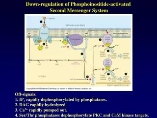
IP3/DAG System The 2nd messenger producing enzyme is phospholipase C. This enzyme hydrolyzes phosphatidyl-inositol- bis-phosphate (PIP2) to produce inositide triphosphate (IP3) and diacyl glycerol (DAG). The enzyme is stimulated by hormones acting through Gi proteins. The PIP2 that is hydrolyzed is produced by phosphorylating the 4 and 5 OH-groups of phosphatidyl inositol using ATP as the phosphate donor. The second important 2nd messenger system that utilizes G-proteins in its transduction mechanism is the one that produces two second messengers upon stimulation by hormones.
Of the two 2nd messengers produced, one is hydrophilic - IP3, and one is hydrophobic - DAG. The effect of DAG is to sequester protein kinase C to the membrane and stimulate its activity. The effect of IP3 is to stimulate Ca++ release from intracellular stores in the SER and mitochondria. The released Ca++ has a variety of effects one of which is the stimulation of protein kinase C.
Protein kinase C is a family of ser/thr protein kinases which include molecules whose activities require stimulation by DAG and Ca++ together, by DAG by itself, , by Ca++ by itself or do not require 2nd messenger stimulation. The first type is the classical protein kinase C, but the other three types have been found by predominantly molecular biological techniques, isolated and characterized.
Gated (K+) Channels The last G-protein modulated 2nd messenger system to be discussed will be ligand-gated K+ channels, neurotransmitter receptors which act to increase potassium conductance in post synaptic neurons act via G-proteins.
Multiple ways that neurotransmitters effect post synaptic cells: There are neurotransmitter receptors which are themselves ion channels (i.e. the ACh receptor), • there are ion channels which are stimulated by neurotransmitter·receptor complexes via G-proteins (i.e. K+ Channels) • and there are neurotransmitters which act through standard 2nd messenger systems (i.e. action of norepinephrine through both and receptors; • receptors are inhibitory to the AC system and receptors are stimulatory to the AC system.
Convergence and Divergence at the Receptor/G-Protein Level A G-protein can be stimulated to exchange its bound nucleotide by a number of hormone·receptor complexes (convergence), and a hormone·receptor complex can interact and stimulate more than one G-protein type (divergence). When one considers the G-protein system overall, one has to realize that there is no one-to-one relationship between a particular G-protein (or G- protein type) and a particular receptor (or receptor type). In addition, a particular G-protein can interact with more than one membrane enzyme, and thus effect more than one signaling pathway (divergence). An example of this is the effect of Gi's on the AC and the IP3/DAG systems.
Tyrosine Protein Kinase Systems Perhaps the most noticeable thing about the tyrosine protein kinase (TPK) system is that there is no "2nd Messenger". The system also uses protein kinases to transmit and amplify signals intracellularly, however the kinase is part of receptor. The binding of hormone by the receptor results in the activation of the tyrosine protein kinase activity. The kinase activity phosphorylates tyrosine residues in substrate proteins, acting in a classical protein kinase stimulatory pathway, thus altering the primary structure and activity of the target proteins. In addition the TKP activity also cross-auto-phosphorylates the receptor/TPK complex.
This autophosphorylation provides a surface for a number of proteins, cys- rich proteins, to bind to. When bound to the membrane surface by receptor/TPK complex, these proteins can catalyze a number of reactions. Among these proteins is phospholipase C, which like other phospholipase C, catalyzes the hydrolysis of PIP2 to yield IP3 and DAG. Therefore, the TPK system is capable of stimulating the second messengers and subsequent pathways of the IP3/DAG system.
A second protein which binds to TPK-P is phosphoinositide-3-kinase which catalyzes the phosphorylation, with ATP as the phosphate donor, of the 3'-OH of inositol residues in phospholipids. This generates a family of 3'-O-P inositides which act to stimulate the Protein Kinase B pathways
Protein Kinase B Phosphoinositide-3-Kinase phosphorylates phosphatidyl-inositides at the 3-OH group. Protein Kinase B binds to the PI-3-P formed And becomes activated by phosphorylation The activate PKB then phosphorylates various substrate proteins, altering their primary structure and thus their function.
The final protein of interest which binds to TPK-P is GRB·Sos which is involved in the activation of the MAP kinase cascade. GRB·Sos acts on Ras, a guanyl nucleotide regulatory protein. In its normal state Ras has GDP bound to a guanyl nucleotide binding site. The presence of GRB·Sos accelerates (causes) the dissociation of GDP from this site, thus allowing it to be replaced by GTP. With GTP in its binding site, Ras is active and activates the MAP kinase cascade.
MAP Kinase Cascade Ras interacts with another protein Raf to form an active ser/thr protein kinase, MAP kinase kinase kinase (MAPKKK), associated with the membrane. The MAPKKK stimulates a protein kinase cascade, through MAPKK and MAPK, which eventually phosphorylates various transcription factors (TF's) and other proteins (Rsk) which also phosphorylate TF's. The resultant TF-P's stimulate the production of new mRNA which in turn leads to the production of new protein and an alteration of cell activity and/or function.
JAK-STAT System The Janus Kinase / Signal Transduction Activators of Transcription system is somewhat similar to the TPK-MAPK system. The binding of ligand causes the receptors to dimerize. JAK binds to the dimerized receptors And phosphorylates them
STAT binds to the phosphorylate receptors And in turn is phosphorylated by JAK; this phosphorylation results in a dimerization and activation of STAT which translocates to the nucleus where it acts as a transcription factor.
Growth Factor – Smed System to form dimeric complex (R-II2•L 2) . The ligand (dimer) initially binds to a high affinity receptor, R-II, This complex recruits a second, receptor (R-I) which has low affinity, to form a R-II2•L 2•R-I2 complex This system is somewhat similar to the last system, however there are two receptors involved and numerous other sites of modulation. The system is the main mechanism by which members of the transforming growth factor ß (TGFß) family of proteins work. These include messengers such as TGFß, bone morphogenic protein, and activin.
R-I is then phosphorylated by a protein kinase which is part of the R-II molecule. The phosyphorylated R-I recruits on of a number of proteins known as SMED’s. Some of these SMED’s are in turn phosphorylated by the kinase activity of R-II.
The phosphorylated SMED’s then dissociates And binds another protein SMED-4 to form a hetero-tetramer, SMED-P2•SMED-42 which translocates to the nucleus and acts as a transcription factor.
Further modulation of the system is accomplished by • Other SMED’s (SMED-6 and SMED-7) which bind to the R-II2•L 2•R-I2 complex but are not phosphorylated by it and thus act as antagonists to standard SMED activation etc. • Other TGFß like proteins such as Inhibins which form complexes with (BG) receptors which in turn recruit R-II receptors to form a larger complex. This complex has no known signaling function and acts to inhibit the activity of the transduction system by tying up R-II in inactive complexes. • Lastly, there are secreted proteins, such as follistatin, which is an Activin binding protein. The follistatin•Activin complex is inactive and is a target for degradation.
Ca+2 systems Calcium is a very important regulator of cell function. It is involved in the control of a large number of motile/fusion process and can act as a second messenger as we have seen already (IP3/DAG system). A variety of cellular proteins act to regulate intracellular Ca+2 levels to include gated Ca+2 channels and Ca+2 Pumps. We have already encountered several gated Ca+2 channels. These include the voltage-gated Ca+2 channel in the synaptic bulb, which allows for Ca+2 entry which in turn leads to vesicle fusion and neurotransmitter release into the synaptic cleft, and the ligand-gated Ca+2 channels of the SER and mitochondria which recognize IP3. In addition there are a variety of 'external' ligand-gated channels which respond to neurotransmitters, and a special channel, the ryanodine channel, which is triggered by Ca+2. The significance of the ryanodine channel is that it allows small changes in Ca+2 concentration to be magnified by release on 'large' intracellular stores. There are two main types of Ca+2 pumps. Those that utilize a Na+ gradient to move Ca+2 and those that use ATP directly.
Calmodulin This small protein binds four Ca+2 ions in a cooperative manner and then interacts with a variety of proteins to alter their activity. The intracellular effects of Ca+2 are manifest through a variety of Ca+2 binding proteins. We have already been introduced to protein kinase C. Perhaps the most important Ca+2 binding protein is Calmodulin.
Among the proteins which calmodulin is known to regulate are adenylyl cyclase, phosphodiesterase, and a variety of Ca+2/calmodulin dependent protein kinases. Other proteins that Ca+2 is known to regulate are phosphoprotein phosphatases, phospholipase A2 and troponin C.
Prostaglandins Phospholipase A2, one of the protein which can be activated by calmodulin, cleaves the fatty acid attached to the 2'-OH of glycerol in phospholipids. This fatty acid is usually arachadonic acid which is a 20- carbon poly-unsaturated (5-6, 8-9, 11-12, 14-15) fatty acid. Subsequent to this cleavage, arachidonic acid is cyclized and oxidized to produce prostaglandins. Prostaglandins generally act as autocrine and paracrine factors. They act on a number of second messenger systems to include the AC system and the IP3/DAG system to augment, control or otherwise modulate the activity of previously stimulated cells or those close by.
Guanylyl Cyclase Systems There are basically two types of guanyl cyclases, one that is membrane bound and has a mechanism analogous to that of the cAMP system and one in which the guanylyl cyclase is soluble. The first type is easy to understand. Hormone binds to receptor and stimulates the activity of guanylyl cyclase, resulting in an increase in the intracellular concentration of cGMP. The cGMP then binds to a cGMP dependent protein kinase (CGDPK) and activates the catalytic activity which in turn phosphorylates protein substrates utilizing ATP as the phosphate donor. The standard 'off' reactions are also present. There is a phosphdiesterase to destroy the generate cGMP and there are phospho-protein phosphatases to dephosphorylate the phosphorylated substrates of the CGDPK. The second system is a little bit more complicated since there has to be a way for the hormone· receptor complex to pass the signal to the soluble guanylyl cyclase. This implies the presence of another (2nd) messenger, and it turns out that this messenger is nitric oxide (NO).
Nitric Oxide (NO) Ligand binding to receptor elevates Ca+2 levels (Ca+2 channel or IP3 mechanism) which stimulates the activity of nitric oxide synthetase (NOSase) resulting in the production of NO from arginine. NO diffuses to soluble guanylyl cyclase and stimulates its activity resulting in the production of cGMP. The increase in intracellular cGMP then acts via standard mechanisms The interesting thing about this mechanism is that NO is lipid soluble and can cross membranes and stimulate neighboring cells. This mechanism means that NO is really an autocrine/paracrine factor rather than being a second messenger.