Download

1 / 26
260 likes | 541 Views
The Second Heart Sound (S 2 ) Chapter 8. Are G. Talking, MD, FACC Instructor Patricia L. Thomas, MBA, RCIS. Components of S 2. Second heart sound The end of ventricular systole & has two high frequency components A 2 & P 2

E N D
The Second Heart Sound (S2)Chapter 8 Are G. Talking, MD, FACC Instructor Patricia L. Thomas, MBA, RCIS
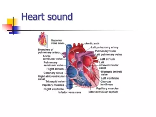
Components of S2 • Second heart sound • The end of ventricular systole & has two high frequency components A2 & P2 • Rapid vibrations of the valves resulting from sudden decrease of blood • 4 factors to determine frequency & intensity of vibrations & affect the production of the aortic and pulmonic components
Components of S2 cont… • The pressure & its rate of development across the closed semilunar valves • The greater the rate of development of the pressure gradient (rapid ventricular relaxation), the more rapid the velocity of valve vibration and the louder the sound produced
Components of S2 cont… • Pliability or stiffness of the value leaflets • Normal or slightly stiff valve leaflets vibrate easily and produce distinct sounds • When heavily calcified reduced mobility as seen in aortic or pulmonic stenosis, they do not vibrate & are associated with decreased or absent A2 & P2
Components of S2 cont… • Valve Radius • Dilatation of the aorta or the pulmonary artery increases their respective valve radius and the intensity of these closing sounds
Components of S2 cont… • Blood Viscosity • Decreased viscosity (anemia) increases the amplitude of these sounds
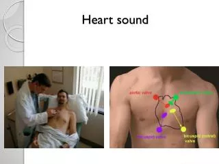
Where to Listen • To hear splitting of S2 • Listen with the diaphragm • Listen with the bell • Second or Third intercostal space, left sternal border • Sitting position, breathing should be quiet, never forced
Physiological Splitting of S2 • Expiration • A2 & P2 are heard as one because of small interval • Inspiration • The splitting of the two sounds is evident • Occurs because increases blood return to the right heart • Increases vascular capacitance of the pulmonary bed • Decreases the blood return to the left heart • Occur in normal children, most young adults, elderly individuals
“Hangout” Interval • Three major intervals of right ventricular contractions are the • Isometric contraction period • Ejection period • “Hangout” interval (a very brief interval at the end of the ejection period & the measure of the impedance of the pulmonary bed) • When impedance is low, the inertia of the blood leaving the right ventricle carries the blood forward even after the force of right ventricular ejection has dissipated
Abnormal Splitting • Wide Splitting of S2 • Audibility of the two components • Paradoxical • Reversed splitting • Reversed in timing (pulmonic closure before aortic closure)
Delay of the Pulmonary Component • Pulmonic Stenosis • Mild Stenosis is a loud sound • Moderate Severe Stenosis unable to hear
Differential Diagnosis • Opening Snap (OS) • Pericardial Knock (PK) • Third Heart Sound (S3) • Late Systolic Click (LSC)
Opening Snap of Mitral Stenosis • Stand patient up to decrease venous return • Any maneuver that decreases LA pressure tend to delay opening snap • Heard in the aortic area
Pericardial Knock • Loud/Sharp and best heard along the lower left sternal border • Constrictive Pericarditis
Absence of P2 • Severe pulmonary stenosis • Single truncal valve • Transposition of the great arteries in children
Pathological Alteration of S2 • Pulmonary Hypertension • P2 louder because of congestive heart failure, mitral stenosis or congenital heart disease • Valve and Vessel Pathology • Diameter and thickening (lacks ability to vibrate in marked thickening and calcification) • Ventricular Dysfunction • Isovolumic relaxation is delayed, the rate of development of pressure across the aortic valve is decreased, produced a diminished A2
THE ENDOFCHAPTER 8 Tilkian, Ara MD Understanding Heart Sounds and Murmurs, Fourth Edition, W.B. Sunders Company. 2002, pp. 72-92.