Download
1 / 1
10 likes | 94 Views
This study investigates the relationship between pain-related fear and lumbar flexion during recovery from low back pain. Participants performed forward bending tasks at different intervals, and joint motions were recorded. Psychological measures like TSK, MPQ, PASS, and PCS were assessed. Results indicate a correlation between pain-related fear and reduced lumbar flexion. Higher pain levels correlated with lesser hip flexion. The study sheds light on how fear influences spine and hip motions during back pain recovery.

E N D
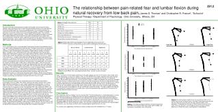
291.2 The relationship between pain-related fear and lumbar flexion during natural recovery from low back pain.James S. Thomas1and Christopher R. France2, 1School of Physical Therapy, 2Department of Psychology, Ohio University, Athens, OH Table 1. Participant Characteristics Introduction Physical examination of low back pain includes the evaluation of trunk range of motion, typically using a trunk flexion task in which the patient bends forward as far as possible with the knees extended and then returns to an upright posture. The examiner observes the relative excursions of the spine and hip while noting any change in symptoms. Although this procedure is a part of the clinical assessment of disability determination, it has not been shown to be closely related to function. Further, while pain-related fear has been associated with avoidance behavior and increased risk for chronic low back pain; few studies have examined how pain-related fear relates specifically to motion of the spine following an acute episode of back pain, and no studies have examined how pain-related fear influences lumbar spine, thoracic spine, and hip motions in this classic examination procedure. Methods Thirty-six participants with a recent episode of low back performed a forward bending task at 3, 6, and 12 weeks following onset of low back pain. Participant characteristics are provided in Table 1. Three-dimensional joint motions of the lumbar spine, thoracic spine, and hip were recorded using an electromagnetic tracking device. Motion Monitor software (Innovation Sports, Chicago, IL) was used to derive time series joint angle data of the thoracic spine, lumbar spine, and hip joints using an Euler angle sequence (flexion, rotation, lateral flexion). Thoracic spine motion was defined by the change in orientation of the sensor on T-1 relative to the sensor on L-1. Lumbar spine motion was defined by the change in orientation of the sensor on L-1 relative to the sensor on the sacrum. Hip motion was defined as the change in orientation of the sensor on the sacrum relative to the sensor on the right thigh. Since the motions used to perform these movements were primarily in the sagittal plane, we restricted our analyses to thoracic spine, lumbar spine, and hip flexion angles. The time series joint angle data were first filtered using a 4th order zero lag butterworth filter with cutoff frequency of 6 Hz. The peak-to-peak changes in joint angles were extracted from the time series data for analyses. Prior to performing the reaching tasks, at each session participants completed the following questionnaires: 1) Tampa Scale for Kinesiophobia (TSK): a 17-item questionnaire that assesses fear of re-injury due to movement, 2) McGill Pain Questionnaire (MPQ):one of the most widely used methods of pain evaluation in both clinical and experimental research, 3) Pain Anxiety Symptoms Scale (PASS):a 40-item measure of pain-related fear, and 4) Pain Catastrophizing Scale (PCS): a 13-item measure describing different thoughts and feelings that individuals may experience when they are in pain. Data Analysis Pearson-correlation analyses were conducted to examine the relationships between psychological measures (MPQ, TSK, PASS, and PCS) and motion (thoracic spine, lumbar spine, and hip) evaluated at 3, 6, and 12 weeks following onset of back pain. As seen in Table 2, there were no significant relationships between the psychological measures and thoracic flexion for any testing session. Kinesiophobia (TSK), pain-related fear (PASS), and pain catastrophizing (PCS) were each associated with less lumbar flexion during a maximum forward bending task at 3 weeks following onset of back pain. Pain-related fear showed the highest correlation to lumbar flexion at each testing session. While lumbar flexion was not significantly related to pain, higher levels of pain were associated with less hip flexion at each follow up interval. To further examine group differences in lumbar spine motion during recovery, participants were assigned to either a high or low pain-related fear group based on a median split of PASS scores from the initial testing session. To assure groups balanced by gender, the median split was conducted separately by sex due to differences in PASS scores for women (Mdn = 56) and men (Mdn = 69). A 3-way MANCOVA for repeated measures design was performed with between subject factors of group (high fear, low fear) and sex and a within subject factor of time from onset of back pain (3, 6, 12 weeks). Back pain ratings from the McGill Pain Questionnaire-Pain Rating Index were included as covariates. Table 2.Relationships between psychological measures obtained at initial assessment (week 3) and joint excursions measured at 3, 6, and 12 weeks following onset of back pain. Note: McGill Pain Questionnaire (MPQ), Tampa Scale for Kinesiophobia (TSK), Pain Anxiety Symptoms Scale (PASS), Pain Catastrophizing Scale (PCS). **p<.001; *p<.05, one-tailed. Results Figure 1 illustrates the effect of pain-related fear on the peak-to-peak excursions of the thoracic spine, lumbar spine, and hip. Specifically, results of the 2 Group x 2 Sex x 3 Time MANCOVA revealed a significant main effect of group (p<.01, ηp2=.23, Observed Power=.82) and a significant group by time interaction (p<.05, ηp2=.19, Observed Power=.59) on lumbar spine excursions. As can be seen in Figure 1 (middle), the group by time interaction reflected a significant increase in lumbar flexion from 3 to 6 weeks in the high fear group (p<.01), but no change between 6 and 12 weeks. Conversely, for the low fear group, lumbar flexion did not change across the three testing sessions. A direct comparison between groups revealed significantly reduced lumbar flexion in the high versus low fear group at 3 weeks (p<.01, one-tailed), 6 weeks (p<.05), and 12 weeks (p<.05). Figure 2 illustrates the effect of pain-related fear on all joint excursions for participants in the low and high pain-related fear groups. This figure suggests that high fear is associated with reduced motion of the lumbar spine, necessitating compensatory adjustments in the joints of the reaching limb and the lower extremities. Conclusion What implications do these findings have for clinical practice? The forward bend task, a standard procedure in clinical examination of the spine, is a composite motion that requires coordination of the hips, lumbar spine and thoracic spine. The relative contribution of these joints in this movement task are often used to help make clinical judgments regarding potential physiologic mechanisms underlying the patients low back pain (e.g. shortened muscles). However, these data suggest that performance of this task is influenced by a combination of pain and fear, each with independent effects on spine and hip joint motions. Thus clinicians should consider the potential role of psychological mechanisms (e.g., fear of pain and harm) when interpreting patient performance on this task. This research was supported by The National Institutes of Health Grant R01-HD045512 to J.S. Thomas Figure 2.The effect of pain-related fear on the maximal excursions of the lumbar spine at each testing session is illustrated. These figures were derived from the mean joint excursions and the mean segment lengths of the participants. The dotted lines illustrate ± 1SD of the mean excursion of the ankle, knee, hip, and spine. The stick-figures clearly illustrate the group differences in lumbar excursions used to perform a forward bend. Figure 1.The effects of group and week on the peak-to-peak joint excursions of the thoracic spine (top), lumbar spine (middle), and hip (bottom) are plotted for each time period.


