Download
1 / 49
490 likes | 499 Views
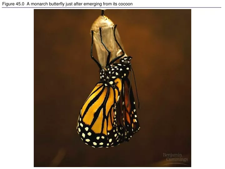
Figure 45.0 A monarch butterfly just after emerging from its cocoon. Figure 45.1 An example of how feedback regulation maintains homeostasis. Figure 45.2 Hormonal regulation of insect development (Layer 1). Figure 45.2 Hormonal regulation of insect development (Layer 2).

E N D
Figure 45.0 A monarch butterfly just after emerging from its cocoon
Figure 45.1 An example of how feedback regulation maintains homeostasis
Figure 45.2 Hormonal regulation of insect development (Layer 1)
Figure 45.2 Hormonal regulation of insect development (Layer 2)
Figure 45.2 Hormonal regulation of insect development (Layer 3)
Figure 11.8 The structure and function of a tyrosine-kinase receptor
Figure 11.10 Steroid hormone interacting with an intracellular receptor
Figure 11.14 The maintenance of calcium ion concentrations in an animal cell
Figure 11.15 Calcium and inositol triphosphate in signaling pathways (Layer 1)
Figure 11.15 Calcium and inositol triphosphate in signaling pathways (Layer 2)
Figure 11.15 Calcium and inositol triphosphate in signaling pathways (Layer 3)
Figure 11.16 Cytoplasmic response to a signal: the stimulation of glycogen breakdown by epinephrine
Figure 11.17 Nuclear response to a signal: the activation of a specific gene by a growth factor
Table 45.1 Major Vertebrate Endocrine Glands and Some of Their Hormones (Hypothalamus–Parathyroid glands)
Table 45.1 Major Vertebrate Endocrine Glands and Some of Their Hormones (Pancreas–Thymus)
Figure 45.6a Hormones of the hypothalamus and pituitary glands
Figure 45.6b Hormones of the hypothalamus and pituitary glands
Figure 45.8 Feedback control loops regulating the secretion of thyroid hormones T3and T4
Figure 45.9 Hormonal control of calcium homeostasis in mammals
Figure 45.10 Glucose homeostasis maintained by insulin and glucagon
Figure 45.11 Derivation of endocrine cells of the adrenal medulla and neurons from neural crest cells
Figure 45.13 Steroid hormones from the adrenal cortex and gonads
Figure 44.24 Hormonal control of the kidney by negative feedback circuits