Download

1 / 99
990 likes | 1.09k Views
Male Reproductive System. Rachel Boggus Boggusrl@email.uc.edu. The Testes. Remember they are located outside the body cavity proper Why? What connects them to the peritoneal cavity? What does this structure consist of?. The Testes.

E N D
Male Reproductive System Rachel Boggus Boggusrl@email.uc.edu
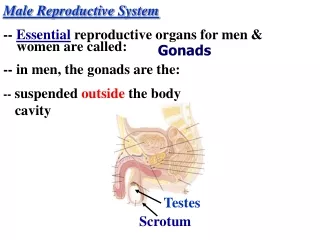
The Testes • Remember they are located outside the body cavity proper • Why? • What connects them to the peritoneal cavity? • What does this structure consist of? Free template from www.brainybetty.com
The Testes • Remember they are located outside the body cavity proper Why? • Lower temperature in testis necessary for spermatogenesis to proceed normally. • What connects them to the peritoneal cavity? • Spermatic cord • What does this structure consist of? • Vas deferens, spermatic artery, pampiniform plexus, and peripheral nerves enclosed by cremaster muscle Free template from www.brainybetty.com
Testes continued • What is the tunica albuginea? • What is contained within the mediastium testis? • What and where is the epididymis? Free template from www.brainybetty.com
Testes continued • What is the tunica albuginea? • Albuginea—collagenous CT capsule • What is contained within the mediastium testis? • Straight tubules, rete testis, efferent ducts • What and where is the epididymis? • Posterior aspect of the testis, highly convoluted where most mature spermatozoa are found Free template from www.brainybetty.com
Seminiferous Tubules • What are the cell types found within them? • What is in the interstitial space? Free template from www.brainybetty.com
Seminiferous Tubules • What are the cell types found within them? • Sertoli cells, and all developmental stages of male gametes • What is in the interstitial space? • Rich vasculature of both circulatory and lymphatic capillaries, myoid cells, and interstitial cells of Leydig Free template from www.brainybetty.com
Note seminiferous tubules Free template from www.brainybetty.com
Interstitial cells of Leydig Free template from www.brainybetty.com
Spermatogenesis • Where does it occur? • How long does it take? • What are the Ad spermatogonia? • What are Ap spermatogonia? • What happens as they increase in size? • What are they called after mitosis? Free template from www.brainybetty.com
Spermatogenesis • Where does it occur? amongst Sertoli cells • How long does it take? 72 days • What are the Ad spermatogonia? Dark, stem cells • What are Ap spermatogonia? Pale • What happens as they increase in size? • Become B spermatogonia • What are they called after mitosis? • Primary spermatocytes Free template from www.brainybetty.com
Spermatogenesis cont. • What do they divide into? • What occurs during this division? • What occurs after 2nd meiotic division? • Conversion of B spermatogoniaearly spermatids is spermatocytogenesis Free template from www.brainybetty.com
Spermatogenesis cont. • What do they divide into? • Two daughter cells called secondary spermatocytes • What occurs during this division? • Autologous recombination • What are they after 2nd meiotic division? • Early spermatids • Conversion of B spermatogoniaearly spermatids is spermatocytogenesis Free template from www.brainybetty.com
spermiogenesis • In this stage they are made to look like tadpoles • 1st stageGolgi stage • Its when the sperm begin to get their heads (acrosomal granule produced) • 2nd stageCap stage • Centrioles migrate to 180º from acrosome • acrosome flattens out • distal centriole gives rise to flagellum • proximal centriole forms connecting piece that joins flagellum with implantation fossa Free template from www.brainybetty.com
Spermiogenesis cont. • Acrosomal stage • most of the acrosome is at anterior pole of the nucleus but thin cap encases the nucleus. Also cytoplasmic microtubules aggretate forming manchette which displacement of most of the cytoplasm forming the residual body Free template from www.brainybetty.com
More spermiogenesis • During acrosomal and maturation stages— • Mitochondria gather around axoneme posterior to nucleus and held in place by hook shaped annulus Free template from www.brainybetty.com
Sertoli cells • AKA nurse cells • Columnar cells with triangular nucleus • Create blood testis barrier (why is this important?) • Shielded from immune system because of recombination making new host cell antigens • REMEMBER THESE JUNCTIONS ARE ABOVE THE LEVEL OF THE SPERMATAGONIA AND THE SPERMATAGONIA ARE NOT IMMUNOLOGICALLY PRIVILEDGED Free template from www.brainybetty.com
Red Arrowssertoli cellsBlack arrowsspermatogoniaBlue ArrowsPrimary spermatocytesGreen Arrowsspermatids Free template from www.brainybetty.com
Blood-Testis barrier • Nutrients must be delivered by RME events facilitated by sertoli cells • Sertoli cells also phagocytize the residual bodies • What hormones do they make and what do these hormones do? Free template from www.brainybetty.com
Blood-Testis barrier • Nutrients must be delivered by RME events facilitated by sertoli cells • Sertoli cells also phagocytize the residual bodies • What hormones do they make and what do these hormones do? • Anti-Mullerian Hormone—inhibits development of the female genital system • Androgen-binding protein—maintains high levels testosterone • Inhibin—negatively regulates (inhibits) gonadotrophsin the anterior pituitary gland Free template from www.brainybetty.com
Interstitial tissue • Leydig cell– what do they look like? • What do they do? • What is this required for? • What are myoid cells? Free template from www.brainybetty.com
Interstitial tissue • Leydig cell– what do they look like? • Large polyhedral cells, usually in clusters and maybe binucleated • What do they do? • Produce testosterone • What is this required for? • Maintenance and function of the accessory glands of male reproductive tract (seminal vesicles, prostate, bulbourethral glands) and the development and maintenance of male secondary sex characteristics • What are myoid cells? • fibroblast (shape) and sm. Musc.—like (contractility) Free template from www.brainybetty.com
Genital Duct System • Where do spermatozoa go after leaving seminiferous tubules? • What are they like? Where do they go? Free template from www.brainybetty.com
Genital Duct System • Where do spermatozoa go after leaving seminiferous tubules? • Straight tubules • What are they like? Where do they go? • Short narrow conduits line with either low columnar or cuboidal simple epithelium • lead to the rete testes Free template from www.brainybetty.com
Straight Tubules Free template from www.brainybetty.com
Genital Duct System • Rete Testis—irregularly shaped tubules located entirely within the mediastinum testis lined with simple cuboidal epithelium Free template from www.brainybetty.com
Rete Testis Free template from www.brainybetty.com
Efferent Ducts • Lined with pseudostratified columnar epithelium what are the 3 cell types? • Goes from testis to epididymis Free template from www.brainybetty.com
Efferent Ducts • Lined with pseudostratified columnar epithelium what are the 3 cell types? • 1) tall columnar ciliated cells • 2)shorter cuboidal cells • 3) basal cells • Combo of tall and short cells gives it undulating appearance Free template from www.brainybetty.com
Efferent Ducts Free template from www.brainybetty.com
Epididymis • What are the 3 regions? • What is the epithelium? • What lines the surface • What is the purpose of the principal cells? Free template from www.brainybetty.com
Epididymis • What are the 3 regions? • Head, body, and tail • What is the epithelium? • Pesudostratified columnar epithelium with stereocilia • What is the purpose of the principal cells? • Main role is absorption of fluid surrounding sperm • Also participates in sperm motility Free template from www.brainybetty.com
Epididymis Free template from www.brainybetty.com
Vas Deferns • Direct continuation of epididymis • Joins prostatic urethra • Epithelium is pseudostratified columnar • Surrounded by three prominent layers smooth muscle Free template from www.brainybetty.com
Vas Deferens Free template from www.brainybetty.com
Efferent = green; seminiferous = black; epididymis = blue Free template from www.brainybetty.com
ACCESSORY GLANDS • START HERE Free template from www.brainybetty.com
The Accessory Glands • Seminal Vesicles—between posterior surface of bladder and the rectum • Highly convoluted • 3 layers—mucosa, muscularis, adventitia • Pseudostratified low columnar or cuboidal cells • “lace-like” appearance Free template from www.brainybetty.com
Seminal vesicles (L) and Prostate (R) Free template from www.brainybetty.com
Seminal Vesicles • Know that they don’t have spermatozoa • Contributes supportive/protective material to the ejaculate Free template from www.brainybetty.com
Prostate gland • Site where male reproductive and excretory systems meet • Has mucosal and submucosal glands • Main prostatic glands in the peripheral regions • Pseudostratified columnar glandular epithelium Free template from www.brainybetty.com
Prostate Free template from www.brainybetty.com
The outer layer is the tunica albuginea and is composed of a dense fibroelastic connective tissue. Note the various profiles of the seminiferous tubules are a result of their convoluted form. Each tubule is ensheathed by myoid cells which have some contractile activity. Free template from www.brainybetty.com
Seminierous – different shapes Free template from www.brainybetty.com
Seminif. tubules on the L, epididymis in upper R Free template from www.brainybetty.com