Download
1 / 79
790 likes | 792 Views
Discover the amazing functions and types of muscles in our bodies, from skeletal muscles that allow us to move to smooth muscles that control organ movements. Learn about the gross and microscopic anatomy of muscles and how they work.

E N D
FREEZE! Don’t move a muscle! • Can you do it? • Nope! • You still breathe, your heart still beats • You can control some muscles, but others work all by themselves!
Muscle Functions • Movement • Maintaining posture and body position • Stabilizing joints • Generating heat • As ATP is used to power muscle contractions, ¾ of the energy escapes as heat, which keeps our body temperature normal • Other functions: • Skeletal muscles protect organs • Smooth muscle is used in valves, arrector pili muscles, and allows pupil to constrict or dilate
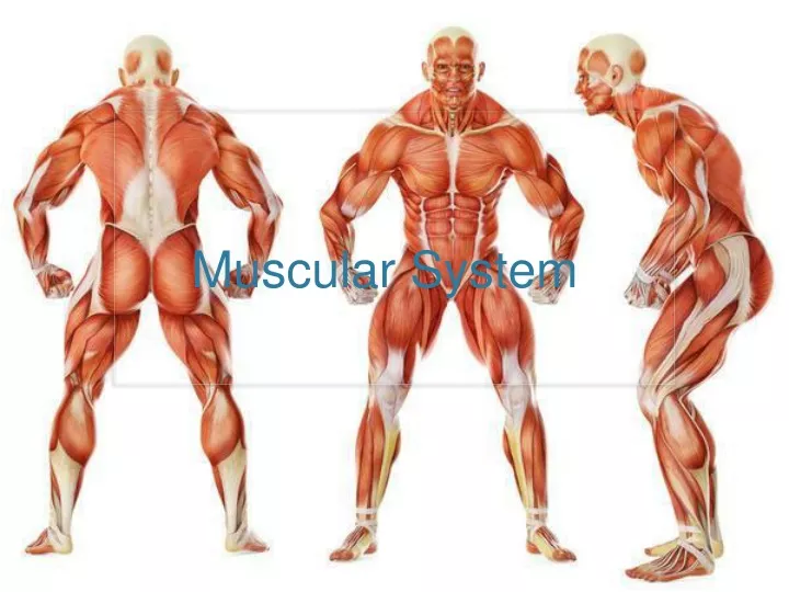
Types of Muscle • Three types of muscle in our bodies: • Skeletal muscle • Smooth muscle • Cardiac muscle
Skeletal Muscle • Voluntary = muscles that are under your control • Ex. Walking, smiling, kicking • Attached to bones, makes bones move • Attached by a tendon, which is connective tissue • Muscle cells are striated • Cells look like they have bands or stripes • Reacts quickly, but gets tired quickly too • Think about a running sprinter
D Gross Anatomy of a Skeletal Muscle A C • Each skeletal muscle is an ORGANmade up of several types of tissue: muscle fibers, blood vessels, NERVEfibers, and CONNECTIVEtissue. E C B
Gross Anatomy of a Skeletal Muscle • The structure of a muscle includes the following parts: • Epimysium = an “overcoat” of dense CONNECTIVEtissue surrounding the whole muscle • Its name means, “UPON the muscle” • Fascicle = each skeletal muscle is made up of bundles that resemble bundles of STICKS
Gross Anatomy of a Skeletal Muscle • Perimysium = fibrous CONNECTIVEtissue that surrounds each fascicle • Its name means, “AROUND the muscle” • Endomysium= within the MUSCLE, each muscle fiber is surrounded by a fine sheath of connective tissue • Its name means, “INSIDE the muscle”
Gross Anatomy of a Skeletal Muscle • In general, each muscle is served by ONEnerve, an artery, and one or more VEINS • Muscles are generally attached to bones at two locations known as the originand the INSERTION
Origins & Insertions of Biceps and Triceps brachii Origins of bicep on scapula Origin of tricep on scapula and humerus Insertion of tricep on ulna Insertion of bicep on radius
MUSCLE ATTACHMENTS • Muscles can be attached either directly or indirectly • Director fleshy attachments = the epimysium of the muscle is fused to the PERIOSTEUMof a bone or perichondrium of CARTILAGE • Indirectattachment = the muscle’s connective tissue wrappings extend beyond the muscle as a ropelike tendon or a sheetlike APONEUROSES • INDIRECT attachments are more common in the body
LABEL THE DIAGRAM: • FASCICLE, TENDON, PERIMYSEUM, EPIMYSEUM FASCICLE PERIMYSIUM TENDON EPIMYSIUM
Smooth Muscle • Involuntary = muscles you cannot consciously control, work automatically • Lines the inside many organs • Ex. Stomach • Control certain movements, especially during digestion • Helps churn food in stomach during digestion • NOT striated • React more slowly and don’t get tired too easily
Smooth Muscle • Cells are spindle-shaped, have only one nucleus, and are surrounded by a thin endomysium • Arranged in two layers • One layer runs circularly, other layer runs longitudinally • Alternately contract and relax to change shape and size of organ
Cardiac Muscle • Involuntary (just like smooth muscle) = automatically works • Found ONLY in your heart • Striated (just like skeletal muscle), looks striped • Does not get tired, muscle is always working • Contractions of cardiac muscle = heartbeats
Cardiac Muscle • Cells are cushioned by a small amount of an endomysium • Muscle fibers are branching cells joined by junctions called intercalated discs • Muscle bundles are arranged in a spiral, which allows the heart to have very coordinated contractions • As the heart contracts, the internal chambers become smaller, forcing the blood into the large arteries
Skeletal Muscle Cells • Each skeletal muscle fiber is a LONG CYLINDRICALcell with multiple oval NUCLEIarranged just below its plasma membrane surface or SARCOLEMMA • Skeletal muscles are huge cells – their diameter is up to 10 Xthe average cell and their length can reach HUNDREDS OF CENTIMETERS long
Microscopic Anatomy of a Muscle Fiber • Sarcoplasm = similar to CYTOPLASMof other cells, but contains unusually large amounts of GLYCOSOMES(stored glycogen) and a unique OXYGEN-BINDING protein called Myoglobin • Myoglobin = a red pigment, similar to hemoglobin, that stores OXYGENwithin the muscle cell
Myofibrils • Myofibrils = rodlike structures that extend the entire length of the muscle cell • Fill up most of the sarcoplasm • HUNDREDS TO THOUSANDSof myofibrils are found in a single muscle fiber • They account for 80%of the cellular volume of the muscle
Striations Come from Myofibrils • Striations come from repeating series of DARK(A) bands and LIGHT(I) bands along the length of each myofibril • A and I bands are nearly perfectly aligned and give the skeletal muscle its BANDEDor striated appearance
I Band and A Band Parts • Z disc = midline interruption of the I band that is darker • H zone = midline interruption of the A band that is lighter • M line = center of the H zone that contains protein rods holding adjacent filaments together • Banding pattern reveals the working structure of myofibrils
Sarcomere • Myofibrils are chains of tiny contractile units called sarcomeres • Sarcomere = the region of the myofibril between two successive Z discs • It is the smallest CONTRACTILEunit of a muscle fiber • Literally means, “muscle SEGMENT”
Myofilaments • Myofilament = small structures within the SARCOMEREthat consist of two types: • Thick Filament-MYOSIN =extends the entire length of the A Band, but does not go into the I band • Helps account for the dark A band and light I band • Thin Filament-ACTIN =runs laterally across the I Band and part way into the A Band • Anchored into the Z disc of the I band • Does not enter the H zone, which accounts for the lighter shade of the H zone
Microscopic Anatomy of a Muscle Fiber ACTIN MYOSIN
Contraction of Muscle Fibers During contraction: • Thin (actin) filaments slide toward each other into the center of the sarcomeres, allowing the actin and myosin to overlap • Makes the light H zone disappear
Sarcoplasmic Reticulum • Sarcoplasmic reticulum = muscle fiber organelle that is a specialized smooth endoplasmic reticulum • Has interconnecting tubules and sacs that surround every myofibril • Like the sleeve of a loosely crocheted sweater • Its purpose is to store and release calcium on demand for when the muscle needs to contract
The Neuron • The neuron, or nerve cell, has many structural parts to it • Axon = long, tail-like extension that sends a message away to another cell • Axon terminals = branched end of the axon that releases the message in chemical form • Neurotransmitter = chemical message • Synaptic cleft = space between an axon terminal and the adjacent cell receiving the message • Terminal NEVER physically touches the other cell x
The Nerve Stimulus • To contract, skeletal muscles must be stimulated by nerve impulses (messages) • Motor unit = one neuron and all the skeletal muscles it stimulates • Axon terminals of the neuron match up to the sarcolemmas of different muscle cells • This creates the neuromuscular junction • Axon terminals will release the neurotransmitter acetylcholine (ACh) into the synaptic cleft when the neuron has been told to do so
The Action Potential • When a nerve impulse reaches the axon terminals: • Calcium channels in the neuron open and calcium (Ca2+) enters the axon terminals • Calcium entry causes the axon terminal to release acetylcholine into the synaptic cleft • Acetylcholine diffuses across the synaptic cleft and attaches to receptors (membrane proteins) within the sarcolemma
The Action Potential • If enough acetylcholine is released, the sarcolemma becomes temporarily permeable to sodium ions (Na+), which come into the muscle cell, and potassium ions (K+) to go out of the cell • More Na+ enters than K+ leaves, which changes the electrical charge of the cell cell is now more positive • Change in electrical charge causes even more Na+ to enter 5. This “upset” generates an electrical current called an action potential • Action potential is unstoppable and it causes a chain reaction throughout the muscle cells, making them contract
After Effects • As long as ACh is present, the action potential will not stop and the muscle will stay contracted • Because of this, ACh must be broken down • Broken down by an enzyme called acetylcholinesterase (AChE) in the synaptic cleft • Produces acetic acid and choline • Once the ACh is broken down, the ion channels can close, the action potential stops, and the muscle can relax
Sliding Filament Theory • We already know that actin slides over myosin during contraction, but how does that actually happen? • Myosin has many “heads” on its ends • When the muscle fibers are activated by the nervous system, the myosin heads attach to binding sites on the actin filaments • Myosin pulls the actin filaments in towards the M line • It is important to note that even though the muscle shortens, the filaments do NOT shorten, they just slide past each other
Importance of Calcium • The attachment of myosin to actin requires calcium ions • Where does the calcium come from? • As the action potential goes further into the cell, it causes the sarcoplasmic reticulum to release calcium ions into the sarcoplasm • Once the action potential ends, the calcium ions are immediately reabsorbed into the SR and the muscle returns to its normal length • This entire process of nerve impulse to calcium reabsorption only takes a few thousandths of a second
3. 4. 2. 1.
The Effect of Exercise on Muscles • Muscles can be changed by the amount of WORK they do or don’t do • For example, when a muscle is used strenuously, it may increase in size or STRENGTH or become more efficient and FATIGUE - resistant • On the other hand, muscle inactivityalways leads to muscle WEAKNESS and wasting
Aerobic Exercise • Aerobic or endurance exercise, such as swimming, JOGGING, and biking result in several recognizable changes in skeletal muscle • These changes in the muscle result in a more efficient metabolism, and in greater endurance, strength, and resistance to fatigue • Changes in the skeletal muscle due to aerobic exercise include: • An increase in the number of capillaries surrounding the muscle fibers • An increase in the number of MITOCHONDRIA within them • An increase in MYOGLOBIN synthesis
Benefits of Aerobic Exercise • Aerobic exercise benefits far more than just the Muscular System. It also benefits other important areas of the body functioning: • Overall body metabolism and NEUROMUSCULAR coordination become more efficient • Improves gastrointestinal mobility (and ELIMINATION) • Enhances the strength of the SKELETON
Benefits of Aerobic Exercise 4. Promotes changes in the cardiovascular and RESPIRATORY Systems that improves the delivery of OXYGEN & NUTRIENTS to the body’s tissues • The heart hypertrophies and develops a greater STROKE volume • Fatty deposits are cleared from BLOOD VESSEL walls • GAS EXCHANGE in the lungs becomes more efficient
Anaerobic Exercise • Anaerobic or resistance exercise, such as WEIGHT LIFTING or isometric exercise causes different changes in the muscle. In these exercises muscles are pitted against high-resistance or IMMOVABLE forces • These changes in the muscle result in muscle HYPERTROPHY, which is an increase in the size of a tissue or organ independent of the body’s general growth • An example of muscle hypertrophy would be the bulging biceps brachii or pectoralis major of a weight lifter
Anaerobic Exercise • Anaerobic or resistance exercise promotes significant increases in the actual SIZE of individual muscle fibers (cells), rather than an increase in the NUMBER of muscle fibers
Muscle Adaptation to Exercise • A vigorously stressed muscle shows increases in the following structures: • MYOFILAMENTS • Myofibrils • MITOCHONDRIA • connective tissue between cells * ALSO STORAGE OF GLYCOGEN INCREASES • However, if the specific exercise routine is discontinued, the muscle fibers will revert to their original metabolic properties
Healthy Muscle Exercise • An exercise program, such as cross training, which alternates AEROBICexercise with anaerobic exercise provides the best program for optimal health • The OVERLOADprinciple applies to both aerobic and anaerobic muscle exercises. • This principle states that forcing a muscle to work hard promotes increased muscle strength andENDURANCE • As muscles adapt to the increased demands, they must be OVERLOADEDeven more to promote further gains
Healthy Muscle Exercise • Because of the overload principle of muscle exercise, it is very important to alternate workout days or conduct a light workout the day following a heavy muscle workout. This will allow the muscles to recover and REPAIRthemselves
Exercise Pitfalls • When healthy exercise principles are not followed, problems may arise • Resistance, or ANAEROBIC, exercise will increase muscle SIZE, but must be done wisely to keep muscles balanced • Because muscles work in ANTAGONISTICpairs (example = biceps brachii and triceps brachii), opposing muscles must be worked equally to maintain balance • When muscle training is not balanced, individuals can become MUSCLE-BOUND. This means they lack FLEXIBILITY, have an awkward stance, and are unable to make full use of their muscles
Exercise Pitfalls • Overuse injuries are common sports injuries that can be caused by the following factors: • Doing too much exercise tooSOON • Ignoring muscle or joint pain
Anabolic Steroids • Anabolic steroids are engineered variants of the natural male sex hormone, TESTOSTERONE • They were first introduced in the 1950s to treat victims of ANEMIA, muscle-wasting diseases, and muscle atrophy (death) in patients immobilized after surgery • Since anabolic steroids are variants of testosterone, they have been shown to increase muscle and bone mass, just as testosterone does in boys during PUBERTY
Risks of Anabolic Steroids • The alleged benefits of steroid use do not outweigh the risks. Risks of anabolic steroids include: • Bloated face • SHRIVELED TESTES • Hair loss • DAMAGE TO LIVER • Infertility • CHANGES IN BLOOD • Raises cholesterol levels • HEART DISEASE * ALSO CAUSES ACNE