Download
1 / 35
350 likes | 519 Views
C-Spine Plain Films. Mike Rissing Associate Student of Clinical Medicine. Outline. Epidemiology Indications for C-spine imaging Modalities Interpretation Types of fractures. Epidemiology. 7000-10000 c-spine injuries treated each year Additional 5000 die at the scene

E N D
C-Spine Plain Films Mike Rissing Associate Student of Clinical Medicine
Outline • Epidemiology • Indications for C-spine imaging • Modalities • Interpretation • Types of fractures
Epidemiology • 7000-10000 c-spine injuries treated each year • Additional 5000 die at the scene • Mean age is 30.7, Mode is 19 • 82% males • 50% MVC, 25% Falls, 10% Sports * www.med-ed.virginia.edu/courses/rad/cspine/index.html
Indications for C-spine Films • Tenderness • Neurologic defecit • Forceful Mechanism of injury • Distracting injury • Altered sensorium
Modalities • Plain films – Lateral, AP, and Odontoid • CT • MRI
Interpretation of Lateral Plain Film • Mnemonic AABCS • Adequacy • Alignment • Bones • Cartilage • Soft Tissue
Interpreting Lateral Plain Film • Adequacy • Should see C7-T1 junction • If not get swimmer’s view or CT
Interpreting lateral Plain Film • Alignment • Anterior vertebral line • Formed by anterior borders of vertebral bodies • Posterior vertebral line • Formed by posterior borders of vertebral bodies • Spino-laminar Line • Formed by the junction of the spinous processes and the laminae • Posterior Spinous Line • Formed by posterior aspect of the spinous processes
Cartilage • Predental Space should be no more than 3 mm in adults and 5 mm in children • Increased distance may indicate fracture of odontoid or transverse ligament injury
Cartilage Cont. • Disc Spaces • Should be uniform • Assess spaces between the spinous processes
Soft tissue • Nasopharyngeal space (C1) - 10 mm (adult) • Retropharyngeal space (C2-C4) - 5-7 mm • Retrotracheal space (C5-C7) - 14 mm (children), 22 mm (adults) • Extremely variable and nonspecific
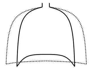
AP C-spine films • Spinous processes should line up. • Disc space should be uniform • Vertebral body height should be uniform. Check for oblique fractures.
Odontoid view • Adequacy: all of the dens and lateral borders of C1 & C2 • Alignment: lateral masses of C1 and C2 • Bone: Inspect dens for lucent fracture lines
Fractures • Mechanisms of injury • hyperflexion i.e. diving in shallow water • axial compression i.e. landing directly on head • Hyperextension i.e. hitting dashboard in MVC
Fractures • Classified as stable or unstable • Stability of cervical spine is provided by two functional vertical columns • Anterior column: vertebral bodies, the disc spaces, the anterior and posterior longitudinal ligaments and annulus fibrosus • Posterior column: pedicles, facets and apophyseal joints, laminar spinous processes and the posterior ligament complex • As long as one column is intact the injury is stable.
Fractures • Jefferson Fracture • Compression fracture of C1 ring • Most common C1 fracture • Unstable • Commonly see increase in predental space on lateral if transverse ligament is damaged and displacement of C1 lateral masses on odontoid. • Obtain CT
Fractures • Burst Fracture • Fracture of C3-C7 from axial loadinng • Spinal cord injury is common from posterior displacement of fragments • Stable if ligaments intact
Fractures • Clay Shoveler’s Fracture • Flexion fracture of spinous process • C7>C6>T1 • stable
Flexion Teardrop fracture • Flexion injury causing a fracture of the anteroinferior portion of the vertebral body • Unstable because usually associated with ligamentous injury
Fractures • Bilateral Facet Dislocation • Flexion injury • Subluxation of dislocated vertebra of greater than ½ the AP diameter of the vertebral body below it • High incidence of spinal cord injury • Extremely unstable
Fractures • Hangman’s Fracture • Extension injury • Bilateral fractures of C2 pedicles (white arrow) • Anterior dislocation of C2 vertebral body secondary to ALL tear (red arrow) • Unstable
Fractures • Odontoid • Complex mechanism of injury • Generally unstable • Type 1 fracture through the tip • rare • Type 2 fracture through the base • Most common • Type 3 fracture through the base and body of axis • Best prognosis
Summary • Know when to order C-spine films: tenderness, forceful injury, altered sensorium, distracting injury, neurologic deficit • Remember your AABC’S • Order CT for evaluation of extent of fracture or MRI if suspect soft tissue injury