Download
1 / 37
430 likes | 490 Views
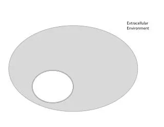
Role of Extracellular Vesicles in Hematological Malignancies. Zahra shakeri niya MSc Student Of Hematology. Cell-to-cell communication. soluble factors Extracellular matrix components ion channels tunneling nanotubules , extracellular vesicles (EV). EV Microvesicle Exosome

E N D
Role of Extracellular Vesicles in Hematological Malignancies Zahra shakeriniya MSc Student Of Hematology
Cell-to-cell communication • soluble factors • Extracellular matrix components • ion channels • tunneling nanotubules, • extracellular vesicles (EV)
EV • Microvesicle • Exosome • Apoptotic body
composition of EV • depending on: • the type cell • physiological and pathological state • proteins • specific lipids • coding and noncoding RNAs • DNA may be present in EV
microenvironmental stressors such as low pH, heat, and oxidative stress modulate the molecular composition of EV
TMV • Microvesicle derived cancer cell named tumor-microvesicle(TMV)
EV cause intracellular communication between cancer cell and microenviroment inhibits physiological function of stromal cells AND promote tumor growth and metastasis.
Tumorigenesis • EV derived from malignant cells affect other cells and change phenotype of normal cells to malignant cell • EV isolated from plasma of B-cell chronic lymphocytic leukemia (CLL) patients can interact and modulate BMSC, thus providing a “homing and nurturing” environment for CLL B cells
Tumor growth • CML derived exosomes are able stimulate bone marrow stromal cells (BMSC) to release IL8, which acts as an in vitro and in vivo prosurvival factor for CML cells. • The inhibition of IL8 receptors, using SB225002, was able to abrogate the IL8- driven CML cell survival in vitro as well as the growth of CML xenograft in vivo.
connect hematopoietic malignant cells and resident cells Prosurvival factor IL8 Activate an AXL mediated pathway CXCLl8/CXCR1-2 signaling
CLL EV maintain the state of activation AKT/mTOR/p70S6K/HIF-1𝛼 modulate the AKT/GSK3𝛽 or AKT/𝛽-catenin signaling pathways Provide tumormicroenvironmentthat favors disease progression
that multiple-myeloma-BM-mesenchymalstromal-cell- (MM-BMSC-) derived exosomes played a role in multiple myeloma (MM) disease progression in vivo. • BMSC transfer exosomes containing miR-15a into MM cells, inducing their proliferation and survival.
Normal neutrophil K562 cells EV • Incubation of neutrophils with K562-EV for 24 hours resulted in the expression of the Bcr/Abl hybrid gene in 20% of the cells • Injection via tail vein of K562 EV into rats or mice caused several symptoms of CML in the animals, such as weakness, loss of weight, splenomegaly, and neutrophilia, but reduced neutrophil phagocytic activity. • Finally transfer BCR/ABL gene with EV to normal neutrophils may promote in vivo transformation of normal cells.
Apoptosis • LAMA84-derived exosomes showed a reduction of the pro-apoptotic genes BAD, BAX and PUMA and an increase in mRNA levels of the anti-apoptotic genes survivin, BCL-xl, and BCL-w.
heat shock proteins (HSP70 and HSP90) and survivinin exosomes cellular stresses inhibit apoptosis and increase cellular proliferation
Angiogenesis • growth, invasion, and metastasis are depending on angiogenesis • Increased angiogenesis in hematologic disorders : AML and CML acute and chronic lymphocytic leukemia MM lymphomas
EV composition changes in target cells or by delivering angiogenic proteins or miRNAs that can stimulate EC function
exosomes released from CML cells directly affect ECs by modulating the process of neovascularization with release of proangiogenic cytokines, such as Interleukin-8
angiogenic miRNAs miR-92 released from K562 enhanced EC migration and tube formation
miR-126 expressed 6-fold more in LAMA84 exosomes compared with the parental cells modulating adhesive and migratory abilities of CML cells
hypoxia boosting tumor angiogenesis with increase of many proangiogenic factors in EV • Hypoxic human leukemia cell line K562 secretes exosomalmiRNAs such as miR-210 higher than normoxic exosomes. suppress the expression of specific genes in endothelial cells, resulting in enhanced pro-angiogenic activity
BM angiogenesis also plays an important role in the pathogenesis and progression of MM • exosome stimulation promotes EC proliferation and the invasion and the secretion of the proangiogenic factors IL6 and VEGF
Tumor immune escape • TMV impairing differentiation and maturation of dendritic cells. • which changed their role from effective APCs into negative modulators of immune response
exosomes enriched in Natural Killer Group 2, member D (NKG2D) thermal and oxidative stress leukemia/lymphoma T- and B-cells Nk cell cytotoxicity NKG2D
Drug resistance • many anticancer drugs can be encapsulated in exosomes and effluxed outside of cells, and shedding of exosomes is closely related to drug resistance in many cancers.
The effect of BMSC-derived exosomes on the survival and drug resistance of MM cells, using both a murine model and human MM samples, has been investigated in MM • drug resistance to bortezomib in MM cells treated with BMSC-derived exosomes, as well as the activation of several pathways related to drug resistance and cell survival, such as NOTCH1, STAT3, NF-𝜅B, and AKT
B-cell lymphoma-derived exosomes carried the protein CD20, exposed in the membrane, able to intercept rituximab, and thus allowing lymphoma cells to escape from humoral immunotherapy • Because exosomal CD20 is a decoy target for rituximab , drug sequestered by circulating exosomes may reduce the efficacy of pharmacological treatment
EV as Biomarkers • EV (both MV and exosomes) contain genetic and proteomic contents that reflect the cell of origin • EV enhancing during cancer progression • EV from body fluids and the circulation of cancer patients is a source of new predictive biomarkers involved in cancer development • EV can protect their cargo from nucleases and proteases, increasing the biomarker half-life
EV-associated cancer biomarkers have been extensively described in a great number of solid cancers, such as prostate cancer, melanoma, and lung cancer • in patients affected with melanoma and other solid tumors, total protein levels of exosome fractions isolated from plasma were reported to reflect disease stage, tumor burden, response to therapy, and survival • in AML, exosomalprotein levels may reflect the Extent of disease and correlate with its relapse after therapy
exosomal protein levels • 1. predictive of long-term disease survival • 2. response to chemotherapy • 3. detect residual leukemia cells in patients considered in complete remission • e.x : the TGF-𝛽1 levels upon AML diagnosis were higher than those in exosomes of normal controls. Following chemotherapy treatment, TGF-𝛽1 levels were significantly reduced, while patients in long-term complete remission had low exosomal TGF-𝛽1 levels
exosomes, released from malignant cells, are enriched in tumor antigens • exosomes isolated from ovarian or breast cancer ascites contain tumor specific antigens HER2/NEU • MART1 is found in nanovesicles recovered from serum of patients with melanoma
mRNAs and miRNAs • EV including mRNAs and miRNAs, to recipient cells, and affect the metabolism of target cells. • That miRNA content from their originating cancer cells is similar to that found in circulating exosomes • Exosomes derived from both AML and CML cells were enriched for several coding and noncoding RNAs relevant to both cancer prognosis and treatment, as well as to the leukemic niche function
miR-155 as a useful biomarker in individuals with monoclonal B-cell lymphocytosis and in patients with B chronic lymphocytic leukemia • higher miR-155 levels expression in patients who failed to respond to chemotherapy compared with those who experienced complete response • At present, miRNA-based clinical trials involving exosomes have not been initiated because an improved characterization of these carriers and their cargos in normal and disease models is still needed
با تشکر از توجه شما The end