Download
1 / 24
250 likes | 485 Views
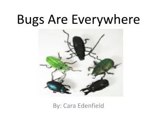
Bacteria are Everywhere . By: Lauren Senter Dr. Hamrick STEP Program at Campbell University. Overview. Part 1 Places we find bacteria Identification Part 2 Control Part 3 Detection in drug products. Places we find bacteria:. Hands Throat Nose Food. Hand Washing Experiment.

E N D
Bacteria are Everywhere By: Lauren Senter Dr. Hamrick STEP Program at Campbell University
Overview • Part 1 • Places we find bacteria • Identification • Part 2 • Control • Part 3 • Detection in drug products
Places we find bacteria: • Hands • Throat • Nose • Food
Hand Washing Experiment • This experiment shows how bacteria normally lives on your skin. When you wash your hands, you remove the surface bacteria that can make you sick but it will not kill or remove all the bacteria on your skin. • To do this experiment: • Make imprint of hand on an agar plate before washing hands. • Then make an imprint of hand after washing hands thoroughly with soap and water. • Incubate until the bacteria grows.
Bacteria in the Throat • For this experiment, we were able to look at and see the different types of bacteria in the throat. • To do this experiment: • Gently rub the back of throat with a sterile cotton swab. • Then rub the bacteria onto a rich media.
Bacteria in the Nose • In this experiment, we were looking for a specific kind of bacteria. • In this experiment: • Using a sterile cotton swab, gently rub the inside of one nostril. • Then rub the bacteria onto an agar plate and incubate for 2 to 3 days. Mannitol Salt agar
Bacteria in Food • In this experiment, we were looking for different types of bacteria in the foods we might eat. • To do this experiment: • Grind up alfalfa sprouts into a fine liquid. • Plate dilutions of the liquid, and incubate to grow up the bacteria. • One of the plates was labeled TNTC (or too numerous to count).The other plate had a total of 165 colonies • 82,500,000 bacteria in a gram of alfalfa sprouts.
Identifying Bacteria • When we find bacteria, there are a number of ways to find out what kinds of bacteria we have.
Gram positive Gram negative rods Cocci (spheres) Cocci (spheres) rods Easy to grow Hard to grow Bacillus subtilis Cells in chains catalase negative Streptococcus pneumoniae E. coli Haemophilus influenzae Cells in clusters catalase positive Staphylococci Flow chart for identifying microorganisms Coagulase positive Staphylococcus aureus Coagulase negative Staphylococcus epidermidis
Identifying Bacteria • Gram Staining is the first step in figuring out what kind of bacteria you are dealing with. • Gram Staining determines whether the bacteria is gram positive, which consists of a cell membrane and a thick cell wall, or gram negative, which consists of an inner membrane, a thinner cell wall, and an outer membrane.
Gram-negative Gram-positive Outer Cell Cell membrane membrane membrane Cytoplasm Cytoplasm Periplasmic space Peptidoglycan Peptidoglycan
1 2 4 3 Don’t Mess Up! • It is important for the bacteria to be a pure culture before putting in a test tube or running any test. • Single colony purification • If you have a mixture of bacteria when you run biochemical test they might appear positive even if some bacteria are negative. • It will cause you to mess up when classifying the bacteria.
Catalase Test • Hydrogen peroxide is a good chemical for killing bacteria. • Catalase is a bacterial enzyme that converts hydrogen peroxide into water and oxygen. • To do this experiment: • Use a loop to remove a little bit of bacteria from the plate and smear it onto a microscope slide. • Add 1 drop of hydrogen peroxide. If the organism produces a catalase, rapid bubbling will occur. • This test helps to recognize if the gram + bacteria is Streptococci or Staphylococci.
Coagulase Test • We did a third test to determine if the bacteria were coagulase positive or coagulase negative. • Coagulase is a bacterial enzyme that clots (or coagulates) plasma products. • This test would tell us if the Gram +, catalase +, bacteria were Staphylococcus aureus or Staphylococcus epidermidis.
Clinical sample Gram positive Gram negative rods Cocci (spheres) Cocci (spheres) rods Easy to grow Bacillus subtilis Hard to grow Cells in chains catalase negative Streptococcus pneumoniae E. coli Haemophilus influenzae Cells in clusters catalase positive Staphylococci Flow chart for identifying microorganisms Coagulase positive Staphylococcus aureus Coagulase negative Staphylococcus epidermidis
Control • Bacteria in the wrong place can make us sick, therefore we have several ways to eliminate or control bacteria. • Some of the ways we can control bacteria are: • Antibiotics • UV light
Antibiotics • Antibiotics kill bacteria. • Different bacteria show different susceptibilities to different antibiotics. • To do this experiment: • Lay little discs with antibiotics in them on plates inoculated with bacteria. • Incubate and you are able to see the different susceptibilities of the bacteria to the medicines. Resistant Sensitive
UV Light • UV light damages bacterial DNA. Different bacteria have different susceptibilities to UV Light. • To do this experiment: • Swab half of each of 4 plates with a spore-forming bacteria, and the other half with a non-spore forming bacteria. • Place one plate (marked 0 min) into the incubator. Place the remaining plates into the hood under the UV light with their lids off. Place the sunglasses over the plate labeled 5min. • Leave the plates in the hood for the allotted time, then take them out and put them into the incubator.
Results • 0min. :All bacteria grew. • 1min. :Bacillus subtilis is able to form spores. Spores are more resistant to UV damage, but some of the bacteria were killed. • Without the ability to produce spores most of the bacteria in the lower part of the plate were killed by the UV light. • 3min. :A little more of the Bacillus continues to die off. The non-spore forming bacteria is almost completely dead. • 5min. :Most of all bacteria are dead except for where the sunglasses were sitting.The sunglasses protected both kinds of bacteria from the UV light.
Detection • To do this experiment: • Grow up Bacillus subtilis as your test bacteria. • Generate a growth curve and add small amounts of bacteria to the media. • The plan was to then add small amounts of Bacillus to a drug preparation and pass through the filter. • Then add media to the filters and wait for the growth of the bacteria.
~117 ~12 ~117 ~12
Steritest • Add drug preparation (containing bacteria) to the filter unit • Add media and incubate • Each filter unit in this test received 2.4 x 10⁴ bacteria.
Sum Up • From doing these experiments, I have learned that bacteria are everywhere and sometimes they belong; sometimes they don’t!