Download
1 / 36
360 likes | 495 Views
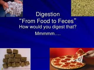
DIGESTION- From sandwich to cells. Darci Barman – Section A. Carly’s Sandwich. Whole-wheat bread Lean turkey Mushrooms Cheese Avocados Tomatoes Mustard/mayonnaise Grapes on the side. Components. Whole wheat bread is often made with: Eggs: provides EAA & cholesterol (+ fats)

E N D
DIGESTION-From sandwich to cells Darci Barman – Section A
Carly’s Sandwich • Whole-wheat bread • Lean turkey • Mushrooms • Cheese • Avocados • Tomatoes • Mustard/mayonnaise • Grapes on the side
Components Whole wheat bread is often made with: Eggs: provides EAA & cholesterol (+ fats) Milk: provides lactose & EAA (+ some fat) Butter: provides saturated fats, cholesterol and some mono- & polyunsaturated fats Yeast: may be a source of trehalose Wheat flour: Provides many EAAs but most likely lacks lysine Provides starch (amylopectin/amylose) Provides cellulose (insoluble fiber) from wheat bran Lean turkey is going to provide the body with all EAA, glycogen and a small amount of fats Mushrooms provide trehalose & some protein & fiber Cheese provides lactose, EAA, saturated fats, mono- and polyunsaturated fats (ratio of these fats depend on types of cheese)
Components Avocados provide a large amount of monounsaturated fats and fiber (3:1 insoluble vs. soluble) Tomatoes although composed mostly of water, provide carbohydrates in the form of starch and soluble fibers Mustard contains a variety of carbohydrates (starch & added simple sugars such as fructose or sucrose), fats (most likely unsaturated) and proteins (will not contain all EAA) – wide variation Mayonnaise is composed of: Eggs: provide all EAA, cholesterol (+fats) Oil: depending on type, contain saturated, poly-and monounsaturated fats Vinegar (acetic acid + water) Grapes will provide fructose & free glucose (+fiber)
Summary of molecules entering digestion • Fats in the form of: • Free fatty acids • TG (containing varying short, medium and long chain FAs of varying saturations) • Phospholipids & cholesterol • Protein in the form of: • Polypeptides – long chains of amino acids (either essential or non-essential) • Carbohydrates in the form of: • Free glucose • Fructose • Lactose (glucose + galactose) • Starch - polysacchardies of glucose – either amylose (linear) or amylopectin (branched) • Glycogen (same as above except from animal sources) • Sucrose • Trehalose • Soluble & insoluble fibers
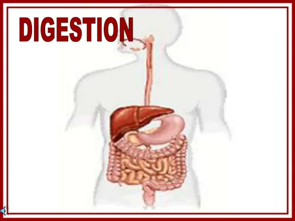
First Stop: The Mouth • The mouth starts the process of digestion by mechanically breaking down and moistening the sandwich components for swallowing with very little chemical digestion (pH = 6.8) • Chewing/presence of food stimulates secretion of saliva to lubricate, provide solvents for interaction with taste buds and to dilute noxious tastes • Saliva also contains enzymes: lingual lipase & salivary amylase • Lingual lipase acts on short & medium chain FAs and hydrolyzes lipids • Salivary amylase is an endoamylase that cleaves alpha-1,4 bonds in starch • Starch Dextrins process begins • No protein digestion occurs in the mouth (large polypeptides remain)
Next Stop: Stomach Station • Mechanical mixing and churning of food in stomach helps emulsification of fats and stimulates G cells to release gastrin • Gastrin released into the bloodstream stimulates parietal cells of stomach to release hydrochloric acid (HCL) • Acetylcholine & GRP (gastric releasing peptide) also stimulate gastric juice release • HCL denatures (uncoils) proteins & coverts pepsinogen to pepsin • Pepsin cleaves polypeptides and inhibits pepsinogen synthesis • Protein polypeptides ---- pepsin, HCL------> smaller polypeptides, some free AA • No carbohydrate digestion occurs in the stomach because high pH (1.2-2) inactivates salivary amylase • Stomach also contains gastric lipase which hydrolyzes TG DG + FA • Very small role overall in fat digestion
Small Intestine, Duodenum • Partially digested food (chyme) enters the duodenum • Chyme stimulates the release of hormones cholecystokinin (CCK) & secretin • Secretin, released from S cells, stimulates the pancreas to release bicarbonate and other neutralizers to protect small intestine from acidic chyme • Optimal pH is 6.9 • CCK stimulates pancreas* and gallbladder* to release various active molecules • Glucose-dependent insulinotropic peptide (GIP) released from K cells to neutralize and slow movement of chyme from stomach to duodenum • *Pancreas releases pancreatic amylase, pancreatic lipase,and proenzymes, cholesterol esterase, and pancreatic phospholipase A2 • *Gallbladder releases bile which contains cholesterol, phospholipid, bile acids, bile salts, electrolytes, antibodies, water & bilirubin (heme byproduct)
Closer look at enzymes released in duodenum • Pancreatic amylase (α-amylase), acts on starches the same as lingual amylase (@ alpha-1,4 glycosidic bonds) • Pancreatic lipase cleaves TG @ carbons 1 & 3 • Bile emulsifies fats to be acted on by lipases • Cholesterol esterase hydrolyzes cholesterol esters to free cholesterols • Phospholipase A2 cleaves phospholipids @ carbon 2 • Proenzymesreleased by pancreas are released in their inactive forms: • Trypsinogen • Chymotrypsinogen • Procarboxypeptidase A • Procarboxypeptidase B • Proelastase • Collagenase • Enzyme enterokinaseactivates trypsinogentrypsin • Trypsin activates other proenzymes, beginning a cascade of enzymatic reactions
Proteases Endopeptidases Exopeptidases Cleave polypeptide bonds at the END of chains (terminal) Carboxypeptidase (active form of procarboxypeptidase) are zinc dependent and cleave at the acid ends A cleaves @ aromatic and branched chain amino acids B cleaves @ terminal arginine & lysine • Cleave polypeptide bonds INSIDE the chain (non-terminal) • Trypsin cleaves peptide bonds @ lysine & arginine • Chymotrypsin (active form of chymotrypsinogen) cleaves peptide bonds @ phe, met, tyr, his, try • Others: elastase & collagenase cleave small polypeptides into di- and tripeptides
BIG PICTURE (duodenum) • Pancreatic amylase (CHO enzyme) • Amylopectin maltose α1,4 + isomaltose α1,6 • Amylose maltose α1,4 • Pancreatic lipases (FAT enzyme) • TG MG (2-monoacylglycerol) + 2 FAs • Isomerase can move fatty acyl to carbon 1 and cleave to free glycerol • Pancreatic phospholipase A2 • PhospholipidLysophospholipid + FA • Pancreatic/intestinal proteases (PRO enzymes) • Polypeptides Di- & Tripeptides and some free AAs • Bile is emulsifying fats from large droplets to small, more accessible droplets
Jejunum & BBM • Jejunum is the segment of small intestine where the majority of macronutrients are absorbed at the brush border membrane (BBM) • Aminopeptidases, dipeptidylaminopeptidasesand tripeptidasesare cleaving the final oligo-, di- & tripeptides left into free amino acids ready for absorption (some di- & tripeptides can be absorbed through BBM) • Location of 5 important disaccharidases (final cleaving into monosaccharides for absorption): • Sucrase: cleaves sucrose to fructose and glucose • Maltase: cleaves maltose to 2 glucose molecules • Lactase: cleaves lactose to galactose and glucose • Isomaltase: cleaves (@α1,6) isomaltose to 2 glucose molecules • Trehalose: cleaves (@ α1,α1) to 2 glucose molecules
Jejunum & BBM (cont) • The last slide indicates that proteins and carbohydrates are ready for absorption as they have been broken down into their respective free amino acids* and monosaccharides but what about fats? • More complicated story… • While being digested in the lumen of the duodenum and jejunum, dietary fats/cholesterols and their products, form “micelles” which are ‘shuttles’ from the site of digestion to the BBM • Micelles contain: • Bile salts • Monoglycerides • Diglycerides • FFA • Lysophospholipids • Sterols
4 Mechanisms of Absorption • Diffusion: the free exchange back and forth through cellular membrane where substances move from a higher concentration to a lower concentration. • Facilitated diffusion: like simple diffusion, it often tries to balance the inner and outer concentrations (high to low) but just needs a carrier protein in order to get through the cell membrane. • Active transport: also requires a protein carrier for substance to move across cell membrane but goes against the concentration gradient therefore takes energy to do so (ATP source of energy, often also requires Na+ involvement) • Pinocytosis: The opposite of endocytosis (substances leaving the cell through membrane wrapped granules). The substance contacts the surface of the cell; the cell membrane wraps itself around the substance (engulfs it), and then releases it inside the cell.
Absorption at BBM • Fats diffuse across BBM in the following forms: • 72% as 2-mono-acylglycerol • 6% as 1-mono-acylglycerol • 22% as free glycerol • FFA diffuse freely • ~50% of dietary cholesterol is absorbed • Amino acids cross the border through active transport • Glucose and galactose are actively transported across BB via SGLUT-1 (sodium-dependent glucose transport 1) • Fructose crosses border via facilitated diffusion (carrier protein = GLUT5)
Who have we left behind? • Left in the intestinal lumen to continue through the digestive system are: • Bile acids not re-absorbed • Nutrients made inaccessible by gel formation (action of soluble fibers) • Minerals, bile acids or lipids adsorbed or bound to soluble fibers • Indigestible soluble and insoluble fibers • Some water…. • The other 50% of dietary cholesterol • Resistant starches
Welcome to the ENTEROCYTE! • Amino acids & glucose may contribute to cell needs otherwise, just passing through • 2-Monoacylglycerols are being re-esterfied into triacylglycerols (monoacylglycerol pathway) • 1-Monoacylglycerols are acted on by intestinal lipase releasing a glycerol and FFA converted to acyl-coA by acyl-coAsynthetase • FFA that entered the enterocyte as so are also acted upon by this enzyme forming a pool of acyl-coA to contribute to re-assembled triacylglycerols (phosphatidic acid pathway) • Lysophospholipids re-esterfied into phospholipids • Cholesterol re-esterfied to cholesterol esters
Lipid Pathways Monoacylglycerol Phosphatidic acid 1-mono-acylglycerol (1MAG) donates glycerol backbone (converted to glycerol-3-phosphate via glycerol kinase) + free acyl-coA fatty acids to form triacylglycerols to be packaged into chylomicrons to exit the enterocyte at BM Other sources of glycerol from glycolysis (oxidation of glucose) • 2-mono-acylglycerol (2MAG) and “acyl-coA pool” build triacylglycerols to be packaged into chylomicrons to exit the enterocyte at BM
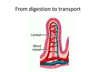
Exit Strategies • Molecules leave the enterocyte at the basement membrane where they enter the bloodstream via the hepatic portal vein (destination: liver) • Glycerol absorbed as so through BBM does not contribute to previously noted ‘lipid pathways’, just “passes through” to portal vein • Amino acids are actively transported to other side* • Glucose, galactose and fructose leave the cell via GLUT2 transporter • Lipids leave the enterocyte as chylomicrons where they are NOT released into the blood, but the lymph • Through the lymph (then, thoracic duct) they will reach muscle & adipose tissue that need the TGs for energy before they are directed to the liver
Chylomicrons • Definition: ‘Ball-shaped’ lipoproteins that contain triglycerides (majority) and cholesterol esters on the inside, surrounded by a phospholipid membrane with cholesterols and apoproteins (specifically, CII & B48) embedded throughout – majority of composition = dietary TGs • On the endothelial cells of capillaries of adipose & muscle tissues resides lipoprotein lipase (LPL) which pulls TG from chylomicrons, taking only the FAs for energy or stored for later use (as TG) • Glycerol backbone released back into the blood liver & kidneys • LPL must be activated by apoprotein CII which also resides on the endothelial cells of the capillary & on chylomicron • After TGs have been pulled from chylomicrons, a high cholesterol-ester containing chylomicron remnant remains to be delivered to the liver
Are We There Yet? YES! The Amazing Liver • The free amino acids from the blood arrive in the liver where they will: • Synthesize liver & plasma proteins (such as albumin) • Be part of free AA pool (in liver OR blood) • Excess will be catabolized: • Amine groupurea* • Carbon skeleton glucoseglycogen*** • Or glucoseacetylcoA fatty acids TG • Pathway that AA follows depends on body’s needs. If the glycogen stores are full, then excess AA takes TG pathway • Glucose enters the liver via GLUT-2 transporters where it has a WORLD of opportunity: • Glycolysis** (for liver energy) • Glycogen*** (energy storage) • Excess acetyl coA TG • Blood glucose • Fructose° and galactose metabolized in the liver (enter via GLUT 2) • Surge in blood glucose stimulates insulin release • CM remnants enter via CM receptor (ApoE-sensitive) by endocytosis
Insulin • After eating this sandwich, the body is in what is known as as FED STATE characterized by high insulin:glucagon ratio. • Insulin stimulates anabolic pathways such as: • Glycogenesis in the liver • BCAA uptake in the muscles for protein synthesis • Glycogenesis in the muscle • TG synthesis in adipose tissue • TG/VLDL synthesis in the liver • Insulin stimulates phosphoproteinphosphatase which DE-phosphorylates (activates) glycogen synthase and (inactivates) phosphorylasekinase which in turn inhibits glycogen phosphorylase (enzyme responsible for freeing glucose from glycogen stores)* • Insulin also stimulates the translocation of GLUT4 receptors to insulin-sensitive cells (adipose, skeletal muscle, cardiac) to increase glucose uptake
Sign, Sealed, Ready for Delivery • The AA & glucose, not directly used for liver function, leave the liver to the tissues • The excess TG previously mentioned either from excess carbohydrates or proteins (+ some cholesterol), is released into the blood via VLDL, a very-low density lipoprotein • Free glucose leaves the liver and is delivered to ALL tissues for normal function (RBCs, retina of the eye & the brain)
Uptake @ Muscle & Adipose Tissue • BCAA are taken up by the muscle tissue for protein synthesis or can be used for energy, whatever the muscle needs at that time… • VLDL arrive and react with LPL (apoCII required) at the capillaries of adipose and muscle tissue • LPL releases FFA for the tissue which it uptakes and uses for energy (muscles, especially cardiac) and stores the rest in the form of TG (for adipose) • The left-over carbon skeletons from TG metabolism head to the liver or kidneys where they can be utilized
Lipoproteins • After the VLDL loose the majority of their TG and apoCII (so LPL cannot degrade any further), it becomes IDL (intermediate-density lipoprotein), also known as VLDL remnant. Since majority of TG have been removed, endogenous cholesterol remains. • IDL can become LDL (low-density lipoprotein) OR go to the liver • If the IDL returns to the liver, it is taken up by the LDL-receptor (which is apoB100 & apoE sensitive), via endocytosis. Both IDL & LDL contain these apoproteins. • As IDL looses more and more TG, it becomes classified as LDL • LDL is taken up into tissues a little differently. The whole lipoprotein is endocytosized and fused with lysosomes inside the cell to break down cellular content.
Lipoproteins (cont) • After LPL fuses with lysosomes, free cholesterol is hydrolyzed (by ACAT) to be used in the plasma membrane or stored intracellularly • Other components of LDL that the cell can use are the glycerol, phosphorus and fatty acids • Although some LDL is taken up in tissues, some is left in the plasma and have a higher chance of being oxidized, phagocytosized and transformed into a foam cell which may contribute to atherosclerosis • The last lipoprotein to mention is HDL, the “good cholesterol” as it’s been coined. It can be generated in the liver or can bud off of other lipoproteins such as chylomicrons and VLDLs when they are degraded by LPL. • They contain mostly phospholipid, some free cholesterol and a variety of apoproteins.
HDL • HDL’s main function is ‘reverse cholesterol transport’. This means that it sweeps free cholesterol from various parts of the periphery and return it to the liver to become biles that is either stored in the gallbladder or sent straight to the GI tract so the cycle can start all over again. • It’s main apoproteins are AI, AII and E • LCAT is a very important enzyme within HDL because it traps the cholesterol in by esterfying it. If it does not, the free cholesterols can just come and go as they please • HDL is taken up by the liver via the HDL-specific, apo-AI sensitive receptor via endocytosis • HDL is considered ‘good’ b/c it pulls cholesterol from the periphery to the liver to be synthesized for bodily needs (ie: bile) therefore reducing the overall cholesterol in periphery AND it impedes LDL oxidation, no FOAM CELLS and atherosclerosis!!
LCAT & CETP • LCAT esterfies free cholesterol to cholesterol esters in HDL • CETP (cholesterol ester transfer protein) is a glycoprotein synthesized by the liver and bound to HDL to transfer cholesterol esters to VLDL and triglycerides and phospholipids to HDL • They both play a critical component in the structure and composition of HDL
Finally! All the tissues look fed and happy. The body is building energy up for the next few hours before another meal
Appendix 1 UREA CYCLE – The conversion of the deaminated AA to urea
Appendix 3 Glygogenesis: Hexokinase above is located in muslce. In the liver, the enzyme is glucokinase.