Download
1 / 62
640 likes | 791 Views
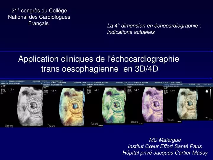
21° congrès du Collège National des Cardiologues Français. La 4° dimension en échocardiographie : indications actuelles. Application cliniques de l’échocardiographie trans oesophagienne en 3D/4D. MC Malergue Institut Cœur Effort Santé Paris Hôpital privé Jacques Cartier Massy.

E N D
21° congrès du Collège National des Cardiologues Français La 4° dimension en échocardiographie : indications actuelles Application cliniques de l’échocardiographie trans oesophagienne en 3D/4D MC Malergue Institut Cœur Effort Santé Paris Hôpital privé Jacques Cartier Massy
Le RT 3DE donne l’erreur la plus petite et les corrélations les plus étroites vs MRI . Sous estimation des volumes , fiabilité de la fraction d’éjection et de la masse JACC 2004
Le matériel • Miniaturisation des sondes matricielles • De la même taille qu’une sonde ETO 2D • Fait 2D ou 3D • Full volume, temps réel, Couleur
Les modalités Temps réel Biplan Biplan 3D temps réel Full volume
Les grandes indications • Anatomie mitrale et annulaire • Procédures interventionnelles: • CIA et foramen ovale perméable • Prothèse aortique percutanée • Fermetures percutanées para prothétiques • AUTRES...;(Tumeurs , th, Vég , Auricule G, etc..)
Prolapsus P2 Grewal JASE 09
Prolapsus bi valvulaire Grewal JASE 09
Perforation valvulaire et fuite para commissurale postérieure
Dilatation mitrale percutanée Perk JASE 09
Commissurotomie mitrale percutanée Eric Brochet Bichat
Mitral valve clopping procedure Perk JASE 09
Plastie mitrale de type Alfiéri Versant ventriculaire Versant auriculaire
Septum atrial : foramen ovale perméable Contraste intra auriculaire gauche
Prothèse Amplatzer : fermeture CIA Vue auriculaire droite
Endocardite aortique Végétation et perforation valvulaire
Athérome aortique Aorte ascendante Aorte descendante